Histology
Evaluation and Interpretation
Evaluation and interpretation is the judgement step that happens after the section is cut, stained and coverslipped: deciding whether what appears down the microscope or on the screen is the patient's tissue or the laboratory's handiwork. A technologist who releases a flawed slide sends a pathologist down the wrong path, so this domain rewards careful looking, pattern recognition and the confidence to hold a case back and recut. It carries roughly 10 percent of the histology paper, but the reasoning it tests runs through every other domain. Evaluation and Interpretation is one of the eight competency areas assessed in the Histology Fields-of-Practice examination set by CAMLPR.
Telling genuine tissue change from procurement and preparation artefact
Almost every slide carries some damage introduced anywhere between the biopsy needle and the coverslip, and the technologist is the first person able to name it. Crush from forceps and cautery damage from the procedure itself, folds, chatter, knife lines, floaters, retraction spaces, stain precipitate, bubbles and freezing holes all mimic pathology convincingly enough to mislead. The habit that separates the two is asking where the feature sits and whether it repeats: anything that runs across tissue and bare glass alike, or lands in the same place on every level, comes from the process rather than the patient. Recognising the pattern lets you name the failing step, recut or restain, and release a slide the pathologist can trust.
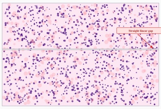
Reviewing H&E levels from a renal core biopsy before releasing the case, a technologist sees a fine straight line crossing every section at exactly the same point, parallel to the direction of cutting. Which conclusion is best supported?
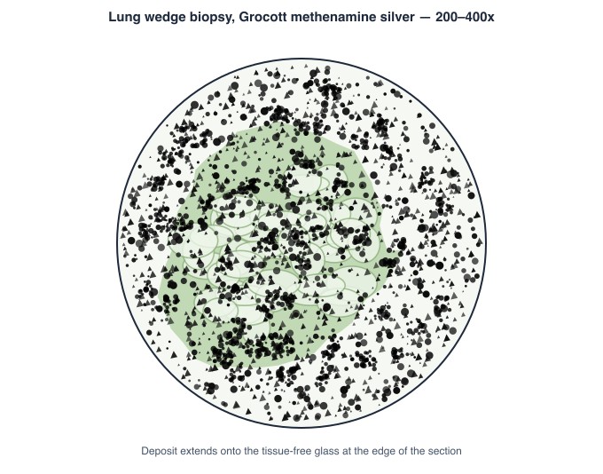
Suspected fungal infection prompts a GMS stain on a wedge of lung tissue. The finished section shows abundant coarse black granular material lying over the tissue and spilling onto the surrounding glass, with no branching or budding forms visible anywhere. What does this black material most likely represent?
Judging scanned slides and digital images before they are reported
Whole-slide scanning moves the quality check onto a monitor, and it introduces failure modes that glass never had. A scan can be perfectly focused at the edges and unusable in the centre, can miss tissue that fell outside the capture area, can tile with visible seams, or can shift colour so that nuclei read brown instead of blue. The technologist reviews the captured image at low and high power before the case is released, compares anything doubtful against the glass slide, and decides whether the fault sits in the section, the stain or the scanner. Only the last of those is fixed by rescanning.
A pathologist reports that a whole-slide scan of a colonic resection is unreadable through the middle of the tissue, although the edges of the same image are crisp at high power. The glass slide is retrieved and checked: the section is flat, intact and properly mounted. What should the technologist do first?
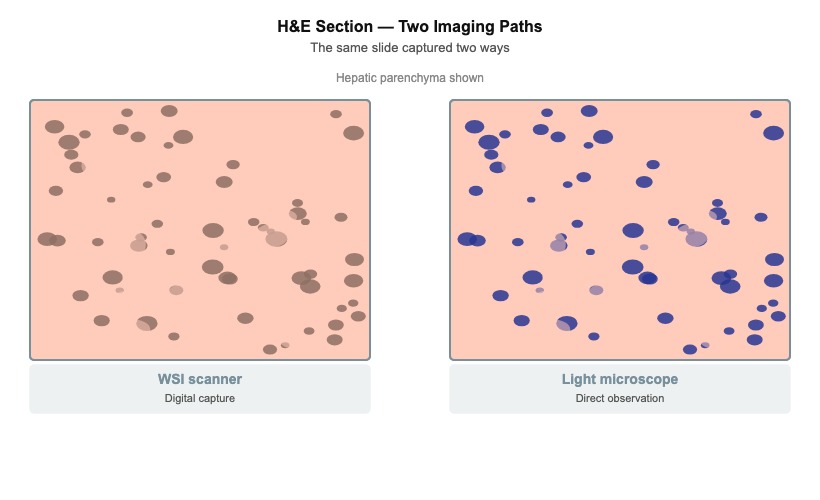
On the reporting workstation, a scanned H&E section of liver shows nuclei that appear brownish-pink instead of the expected blue-purple, and the whole image carries a warm cast. The same slide examined down the microscope shows normal nuclear and cytoplasmic colour. What is the most likely explanation?
Keep reading with a free account
The rest of this page — 1 more section and 2 practice questions — is free to read with an MLTPrep account.
No card required.
Back to all histology areas in the Histology study guide.
MLTPrep is an independent study resource. Not affiliated with or endorsed by CAMLPR. Always confirm current requirements with CAMLPR directly.