Clinical Chemistry
Preparation and Testing
Preparation and testing is the analytical core of clinical chemistry — everything between a specimen arriving fit for use and a number appearing on the screen. It covers identity checks at each transfer, reagent and control preparation, microscopy of urine and body fluids, molecular set-up, blood gas handling, and the separation techniques a laboratory runs. It also covers knowing where each method stops being reliable, because a technologist who cannot recognise an interfered result will release it. Preparation and Testing is one of the eight competency areas assessed in the Clinical Chemistry Fields-of-Practice examination set by CAMLPR.
Keeping the specimen tied to the right patient
Every transfer is a chance to break the link between a tube and the person it came from. You confirm that the name and unique identifier on the tube agree with the requisition when the specimen is received, again when it is aliquoted, again when it is loaded onto an analyser, and once more if it is stored or sent away. Aliquots carry their own full labels, never a rack position or a sequence number. Where tube and paperwork disagree, the specimen is not tested and not relabelled — it is rejected and recollected, with the discrepancy documented.
A technologist decants serum from one primary tube into three aliquot tubes for send-out testing at a referral centre. The primary tube carries a complete patient label. What must be done before the aliquots leave the bench?
A lithium heparin tube arrives labelled "J. Okafor" while the requisition accompanying it names "J. Okonkwo". Both patients are inpatients on the same ward. What is the correct action?
Making up reagents, calibrators, standards and controls
Assay performance is decided before the first sample is loaded. This area covers reconstituting lyophilised material with the stated diluent and volume, allowing it to dissolve fully, mixing gently rather than shaking, and labelling every working solution with its preparation date, expiry and preparer. You track lot numbers, honour open-vial and on-board stability, store material at the temperature the insert specifies, and recalibrate when a reagent lot changes rather than carrying an old curve forward. Small volumetric errors here are multiplied into every patient result that follows, which is why the arithmetic is checked rather than trusted.
A chemistry analyser is loaded with reagent drawn from a fresh enzymatic creatinine lot. The calibration from the previous lot is still inside its stated stability window. What must happen before patient results are reported on the new lot?
Lyophilised multi-analyte control material is reconstituted at the bench with a volumetric pipette, swirled and left to dissolve as directed. Quality control for every enzyme in the panel then falls above the assigned range. Which preparation fault best explains this?
Recognising what is in the field down the microscope
Chemistry microscopy is largely urine and body fluid work, and the first task is naming what you see. You separate cells from crystals from casts from artefact, and you read each finding against everything else known about the specimen — the pH, the specific gravity, the reagent strip results, the clinical note. A crystal that cannot form at the recorded pH has probably been misidentified. You also know which elements are reportable, which are incidental, and which need referral, and you confirm anything unusual at higher power before it is entered.
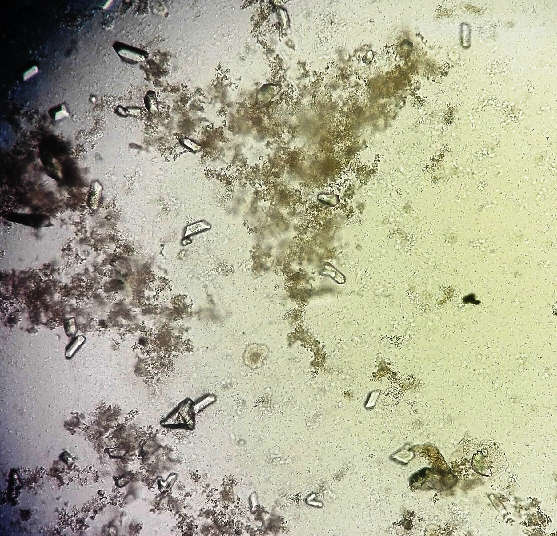
Urine sediment from a patient being investigated for recurrent infection is examined at 400x [IMAGE]. The reagent strip on the same specimen recorded a pH of 8.5. Which crystal is consistent with both the morphology shown and that pH?
Knowing where your method stops being reliable
No assay is right for every specimen, and this area asks whether the result in front of you falls inside what the method can actually do. You know the analytical measuring range and what happens above it, the interferents each chemistry is vulnerable to — icterus, haemolysis, lipaemia, drugs, paraproteins — and the structural limits such as the dilution step in an indirect ion-selective electrode or the non-specific chromogens of an alkaline picrate reaction. When two methods disagree, you can say which one the specimen has broken, and choose an alternative rather than reporting a plausible-looking number.
A serum sodium of 122 mmol/L is produced by the indirect ion-selective electrode on the main chemistry analyser. The patient is clinically well and the specimen is grossly lipaemic. A blood gas analyser using a direct electrode gives 139 mmol/L on the same draw. What explains the difference?
A laboratory measures creatinine by the kinetic alkaline picrate (Jaffe) reaction. Which specimen is most likely to give a falsely raised creatinine by this method when compared with an enzymatic assay?
Preparing slides that can actually be read
A poor preparation cannot be rescued at the microscope. This area covers choosing the right technique for the specimen and the cell count in front of you — a direct wet preparation, a stained smear, or a cytocentrifuge spin when a body fluid is too dilute for cells to be found on a plain film. You control the volume applied, the coverslip, the drying and the stain, you work promptly before cells degenerate, and you label each slide so that it stays linked to its specimen through staining, reading and storage.
A cerebrospinal fluid specimen with a nucleated cell count of 12 × 10⁶/L is submitted for a differential. Which preparation gives the best cellular detail for that differential?
Setting up amplification reactions without contaminating them
Molecular work in a chemistry department demands a discipline the rest of the bench does not. You extract nucleic acid, assemble master mix and template in a defined order, and include the controls the assay requires: a no-template control, a positive control, and an internal control inside every reaction. Movement through the workspace runs one way only, from clean reagent preparation to specimen addition to amplification, with dedicated pipettes, filter tips, coats and equipment in each area. A single previous amplicon carried backwards is enough to make an entire run unreportable.
A molecular bench in a chemistry department finds intermittent low-level positives in its no-template controls across several runs, with no other change to reagents or staffing. Which working practice most directly addresses the likely cause?
A qualitative PCR assay returns no signal for the target and no signal for the internal control in one patient specimen, while the run's positive and negative controls behave exactly as expected. How should this specimen be reported?
Judging whether the specimen is fit to test
Before anything is analysed the specimen itself is assessed. You check that the tube type matches the request, that the fill reaches the stated draw volume, that an anticoagulated specimen is free of clots, and that enough serum or plasma exists for the tests ordered plus a repeat. You check timing and transport conditions for analytes that need them, and you look at the specimen — its colour, its turbidity, any layering. Where a specimen fails, you document why, tell the requester, and ask for a replacement rather than testing something already known to be compromised.
A lithium heparin tube sent for electrolytes contains a visible fibrin clot after centrifugation and yields only a small plasma layer. The potassium returns 6.8 mmol/L on a patient with no clinical features of hyperkalaemia. What is the correct next step?
A 24-hour urine for creatinine clearance is delivered with a recorded volume of 400 mL and a note from the patient saying one overnight void was passed into the toilet. What should the laboratory do?
Examining body fluid cells and the material around them
Beyond urine, chemistry laboratories examine cerebrospinal, synovial, pleural, peritoneal and pericardial fluids. This area covers performing chamber counts and calculating correctly from the area and depth actually used, distinguishing cell types on a stained preparation, and recognising non-cellular material such as crystals, fibrin, fat globules or contaminating debris. Crystal identification under compensated polarised light — shape, strength of birefringence and its sign — is a distinct skill with real clinical consequences. Fluids are handled quickly, because their cells break down within hours of collection.
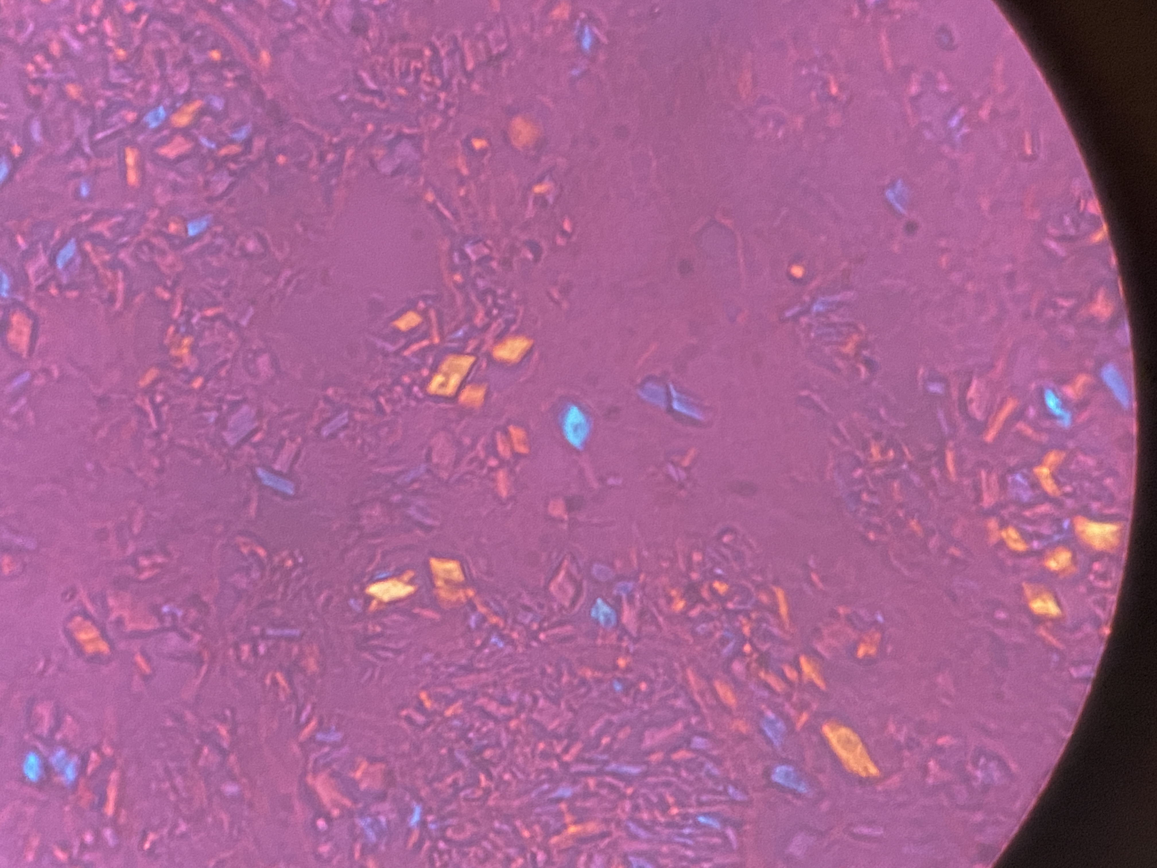
Synovial fluid from a swollen knee in a 78-year-old is examined under compensated polarised light [IMAGE]. The crystals are rhomboid and short-rodded, weakly birefringent, and appear blue when their long axis lies parallel to the compensator's slow axis. Which crystal is present?
An undiluted cerebrospinal fluid is loaded into a Neubauer chamber for a nucleated cell count. The technologist counts 60 cells across all nine large squares on one side of the chamber. What is the nucleated cell count?
Separating a mixture, then measuring the parts
Separation starts at the centrifuge: spinning a specimen into serum, plasma or cells at the speed and time the method requires is the everyday case. Beyond that, some analytes have to be pulled apart from one another before they can be measured at all. This area covers the separation techniques a chemistry laboratory runs — zone electrophoresis, immunofixation, high performance liquid chromatography, and the mass spectrometry systems layered on top of them — and what each one is separating by. You know that electrophoresis on cellulose acetate sorts proteins by net charge at the running pH, that a chromatographic peak is defined by its retention time, and that an unexpected extra band or peak is information rather than noise.
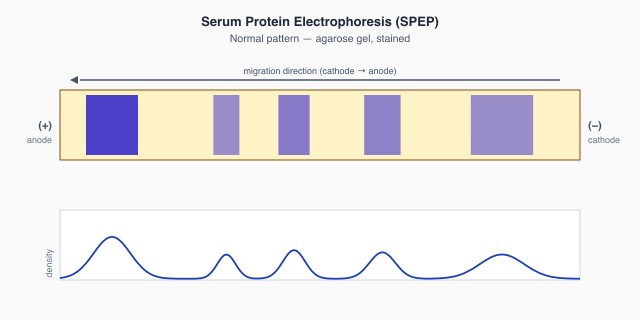
A serum protein electrophoresis is run on an agarose gel in a barbital buffer at pH 8.6 [IMAGE]. Which property of a protein determines how far it migrates from the point of application?
Haemoglobin A1c is measured by cation-exchange HPLC. One patient's chromatogram carries a large unidentified peak eluting after the A1c peak, and the reported A1c is implausibly low against the recorded glucose history. What should the technologist suspect?
Putting a number on what the microscope shows
Reporting "some" or "a few" is not enough; findings have to be graded the same way between technologists and between shifts. This area covers counting elements per low power or per high power field according to what the element is, using chamber counts where an absolute number is required, and applying the semi-quantitative grading scale the laboratory has defined. The concentration factor matters — volume centrifuged divided by residue retained — because changing it changes every count per field and invalidates the ranges built on the original method.
A laboratory changes its urine sediment protocol from centrifuging 12 mL to centrifuging 6 mL, while keeping the same 1 mL residue, the same drop size and the same coverslip. What happens to the number of elements counted per field?
Reading urine by eye and by reagent strip
The physical and chemical examination of urine is quick, cheap and easy to get wrong. You describe colour and clarity against a standard vocabulary, measure specific gravity, and read each reagent strip pad at its own stated time under good light or on a reflectance reader. Just as important is knowing what each pad actually detects and what defeats it — the protein pad's bias toward albumin, ascorbic acid quenching blood and glucose, alkaline urine producing a false protein reaction, delay changing everything. Discrepancies between pads, or between a pad and the sediment, are followed up rather than reported.
A patient with known multiple myeloma has a urine reagent strip protein pad reading of negative, while a sulphosalicylic acid precipitation test on the same specimen is strongly positive. What accounts for this?
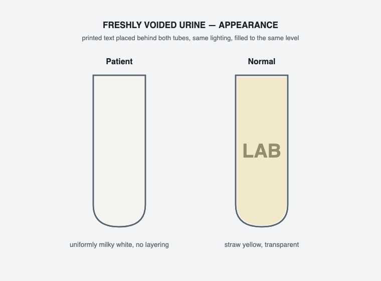
A freshly voided urine is uniformly milky white and opaque throughout [IMAGE]. The turbidity does not clear on warming or on acidification, and the reagent strip shows a strongly positive leucocyte esterase with a positive nitrite. Which explanation best fits?
Handling and running blood gas specimens
Blood gases are the most pre-analytically fragile work in the laboratory. You check that the syringe is anticoagulated correctly and not over-heparinised, that no air bubble remains, that the specimen is identified with its collection time and the patient's oxygen therapy, and that it reaches the analyser within minutes rather than sitting on a bench. You mix thoroughly before aspiration, you know how ongoing cell metabolism drives pO₂ and glucose down while pCO₂ climbs, and you can separate a genuine acid-base disturbance from an artefact created in transit.
An arterial blood gas syringe is drawn correctly, capped and mixed, but then sits at room temperature for 35 minutes before it reaches the analyser. Which pattern of change is expected in the reported results?
A blood gas syringe arrives holding a 0.5 mL air bubble that has been mixed through the specimen during transport by pneumatic tube. The patient is breathing room air and is known to be hypoxaemic. Which result is most affected?
Working through the urine sediment
Microscopic examination follows a set routine: a standard volume centrifuged at a standard speed, a standard residue resuspended, a set drop under a coverslip, casts and larger structures scanned at low power with the light reduced for contrast, then cells, crystals and organisms confirmed at high power. You know why hyaline casts vanish under bright illumination, why a cast localises a finding to the nephron rather than the lower tract, and why a specimen left standing for hours grows bacteria, shifts pH and loses the fragile elements you were looking for.
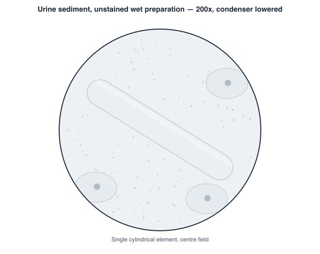
A urine sediment is examined with the condenser lowered and the light reduced [IMAGE]. Why is this illumination used when searching for casts?
Red cell casts are seen at 3 per low power field in the sediment of a patient presenting with visible haematuria and new hypertension. What does the presence of the casts establish?
Spotting haemolysed, lipaemic and icteric specimens
Three visible specimen states change results, and this area asks you to recognise them, know which tests they hit, and decide what to do. Haemolysis floods the plasma with potassium, lactate dehydrogenase and aspartate aminotransferase from inside the red cells. Lipaemia scatters light in photometric assays and, through volume displacement, pulls an indirect sodium down. Icterus interferes with reactions read in the same wavelength region as bilirubin. You use the analyser's indices rather than the eye alone, apply the documented rejection and comment rules, and where a specimen can be salvaged — ultracentrifugation for lipaemia — you do that instead of reporting a biased number.
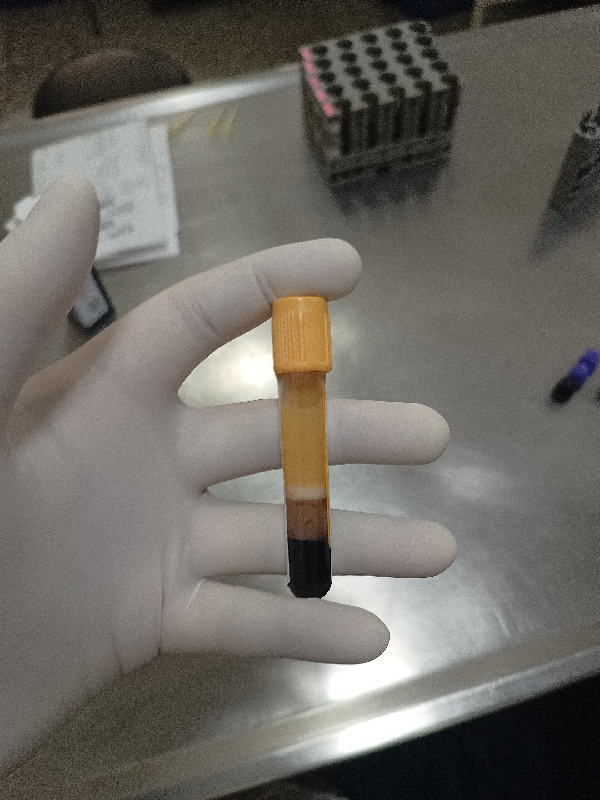
A fasting serum for a routine chemistry profile is grossly turbid after centrifugation [IMAGE]. The analyser raises a lipaemic index flag and the sodium is reported as 128 mmol/L on an indirect electrode. Which handling step best allows a valid sodium to be issued?
A serum specimen sent for electrolytes is visibly pink after centrifugation, and the analyser reports a haemolysis index of 3+. Which analyte is changed most by that haemolysis, and in which direction?
Keep reading with a free account
The rest of this page — 13 more sections and 23 practice questions — is free to read with an MLTPrep account.
No card required.
Back to all clinical chemistry areas in the Clinical Chemistry study guide.
MLTPrep is an independent study resource. Not affiliated with or endorsed by CAMLPR. Always confirm current requirements with CAMLPR directly.