Histology
Specimen Collection
Histology rarely gets a second chance at a specimen. Tissue is taken once, in an operating theatre or a clinic room, and everything that follows — fixation, embedding, staining, diagnosis — inherits whatever was done in those first minutes. This domain covers the checks, containers, labels, transport and storage decisions that decide whether a biopsy arrives at the cutting bench identifiable, intact and fit to report. Specimen Collection is one of the eight competency areas assessed in the Histology Fields-of-Practice examination set by CAMLPR.
Checking the requisition before anything else happens
Every tissue specimen travels with paperwork, and HIST-SC-1 covers the checks made against that paperwork before a case is accessioned. The technologist confirms that the identifiers on the container and on the requisition agree, that the anatomical site and the side are both stated, that the responsible clinician and the collection date and time are recorded, and that the clinical summary supports what has been requested. Histology adds a further layer: the requisition signals whether unfixed tissue must be triaged for flow cytometry, microbiology, electron microscopy or a frozen section. A mismatch is a hold point, resolved with the ward rather than guessed at.
A skin ellipse reaches the accessioning bench in a dermatology batch. The requisition names the left forearm as the excision site; the container label reads right forearm. Nothing else differs. What is the correct next step?
A neck lymph node arrives in the cut-up room during the afternoon list, and the accompanying requisition raises the possibility of lymphoma. Which entry on that requisition most directly changes how the tissue is handled on arrival?
Getting the patient ready for a tissue procedure
Tissue collection is a procedure performed on a person, so HIST-SC-2 covers the preparation that happens before a needle or blade is used. Documented consent must be present. Allergies to local anaesthetic, latex and skin antiseptic are checked. Anticoagulant and antiplatelet therapy is held according to local protocol, and recent coagulation and platelet results are available for core and marrow biopsies. The patient is positioned for access and comfort, told what to expect and how long it will take, and privacy is arranged. Afterwards, pressure, dressing and observation instructions are given. Laboratory staff supporting these lists are expected to confirm each item rather than assume the clinic did it.
A technologist is assisting at an ultrasound-guided renal core biopsy list. The first booking is an adult on long-term warfarin for atrial fibrillation. Which item must be confirmed before the procedure begins?
Proving the specimen belongs to the right person
HIST-SC-3 covers positive identification at the moment tissue is taken. Two unique identifiers are matched between the person, the wristband or chart, and the requisition, and the container is labelled there and then rather than later at a bench. Where the patient cannot take part — under anaesthetic, unconscious, very young, or confused — a second qualified staff member or an accompanying adult verifies instead. An unidentified emergency admission is handled under the assigned temporary identifier, which is later reconciled to the permanent record. Surnames repeat, room numbers get reassigned, and clinic lists run out of order, so none of those may stand in for a unique identifier.
An unresponsive trauma admission with no name on file is taken to theatre, where a wedge of devitalised muscle is excised for histology. How should identification be handled for that container?
Two people on a dermatology clinic list share the surname Nwosu and are booked minutes apart, both for shave biopsies of pigmented lesions. What best protects against a specimen mix-up at the point of collection?
Choosing the right container and collection tools
HIST-SC-4 covers matching hardware to the tissue and to the investigation requested. Containers must be leak-proof, wide-mouthed enough that a fixed specimen can come back out, and large enough for the volume of fixative required. Very small fragments need a biopsy cassette lined with foam pads or a fine mesh bag so they are not lost in processing. Specialised work needs specialised vessels: a glutaraldehyde vial for electron microscopy, transport fluid for direct immunofluorescence, sterile culture medium for flow cytometry, a dry sterile container when microbiology must sample first. Choosing wrongly at collection cannot be corrected once the tissue is in the wrong liquid.
An endoscopy unit is sending several duodenal fragments, each about two millimetres across, for coeliac disease investigation. What should be supplied so the fragments are not lost between collection and embedding?
A thoracic surgeon plans a lung wedge resection on a patient with a cavitating lesion, and asks for both mycobacterial culture and routine histology on the same piece of tissue. What should the operating theatre be supplied with?
Having fixative and supplies ready before the list starts
HIST-SC-5 covers preparing the consumables that collection depends on. Containers are pre-filled with enough 10% neutral buffered formalin to give roughly ten times the volume of the expected specimen, because underfilled fixative is the root of most poorly fixed large resections. Lot numbers and expiry dates are checked on fixatives and transport media, and stock is rotated. Decanting from bulk drums happens under local exhaust ventilation with gloves, gown and eye protection, and every decanted container leaves the laboratory with a workplace hazard label naming the contents and the hazard. Cassettes, foam pads, inks, forceps and sterile containers are stocked ahead of the list.
A theatre nurse telephones before a list to ask how much 10% neutral buffered formalin should already be in the container for a specimen expected to be about 50 mL in volume. What volume should be supplied?
A technologist decants 10% neutral buffered formalin from a bulk drum into small screw-cap containers under the fume hood, ready for a visiting dermatology clinic. What must be done before those containers leave the laboratory?
Labelling that survives formalin and scrutiny
HIST-SC-6 covers writing identity onto the specimen itself. Labelling happens at the point of collection, in the patient's presence, never in advance and never afterwards from memory. The label goes on the body of the container, not on the lid, and carries at least two unique patient identifiers plus the anatomical site, the side, and the collection date and time. Ink and label stock must resist formalin, alcohol and xylene, since a legend that dissolves in the processor is no legend at all. Where several specimens come from one session they are numbered in a sequence that matches the requisition exactly, site by site.
A set of prostate cores arrives with the patient identifiers written neatly on the lid of the container and nowhere else. Why is that practice rejected in histology laboratories?
A colonoscopy performed for surveillance yields biopsies from four separate levels of the large bowel during a single session. How should the containers be labelled?
Moving specimens without damaging or spilling them
HIST-SC-7 covers what happens between the collection room and the laboratory. Fixed specimens travel in a sealed leak-proof container inside secondary containment, usually a biohazard bag with the requisition in an outer pocket so paperwork stays dry and readable. Formalin in transit between sites is subject to dangerous goods packaging rules, and bulky containers are usually excluded from pneumatic tube systems. Unfixed tissue is a different problem: it must not dry out and must not be frozen if paraffin sections are wanted, so it travels on saline-dampened gauze, chilled, and quickly. Medico-legal specimens travel under a documented chain of custody.
A margin from a breast lumpectomy must go from the operating theatre to the laboratory for an intraoperative frozen section, a journey of about five minutes. How should the tissue be sent?
A satellite clinic 40 kilometres from the main laboratory sends fixed skin biopsies by daily courier. What packaging requirement applies to the formalin-filled containers for that journey?
Judging whether the specimen is fit to report
HIST-SC-8 covers the assessment made when tissue is received and again when it is grossed. The technologist looks at whether the specimen matches what the requisition describes, whether it is adequate in size for the question asked, whether it is fully covered by fixative, and how long it sat before fixation began. Histology has its own failure modes: crushing from forceps or biopsy jaws, drying at the edges, autolysis in the centre of a large unincised resection, freezing artefact in tissue that was chilled too hard, and cautery damage along a diathermy margin. Unsuitable specimens are documented and discussed, not quietly processed.
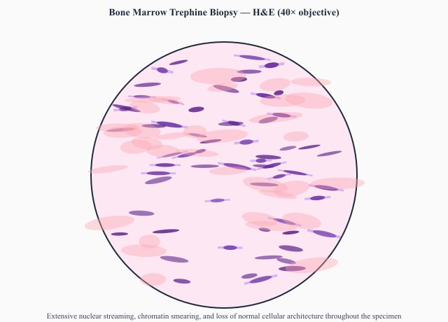
A haematology team submits a trephine core taken during a marrow procedure on a 62-year-old under investigation for a myeloproliferative disorder. [IMAGE] shows nuclear streaming and smeared chromatin through much of the core. Which event during collection best explains this appearance?
A hysterectomy specimen is placed intact into a container of formalin late on a Friday and reaches the laboratory on Monday morning. At cut-up the outer myometrium looks firm and pale, while the deep myometrium is soft, dull and grey. What accounts for the deep change?
Triage and accessioning at the receiving bench
HIST-SC-9 covers turning a received container into a live laboratory case. The specimen is accessioned against a unique case number, the requisition data are entered, and the number is written onto the container so tissue and record cannot separate. Where more than one investigation is requested, the tissue is divided at this point according to a written allocation protocol, because each destination needs a different medium and the split cannot be reversed. Time of receipt and time into fixative are recorded, since fixation duration is later needed to interpret immunohistochemistry. Urgent, rapid and intraoperative cases are flagged and routed ahead of the routine batch.
A renal needle-core biopsy is divided in the laboratory between light microscopy, direct immunofluorescence and electron microscopy. Into which medium does the fragment reserved for direct immunofluorescence go?
Storing tissue, blocks and slides after reporting
HIST-SC-10 covers where material lives once testing is under way and after the report is signed. Residual fixed tissue is kept in a wet-tissue store that is ventilated for formaldehyde vapour, secured against casual access, and organised so any case can be retrieved on request. Blocks and slides are archived dry, at stable room temperature, away from heat and direct light, for the retention period set by the laboratory's own quality programme and provincial rules. Unfixed material held for molecular or research work goes to a monitored ultra-low freezer with temperature logging and alarms. Retrieval, disposal and destruction are all recorded.
A small remnant of unfixed tumour left over from an intraoperative frozen section is to be kept in case molecular testing is requested weeks later. How should that remnant be stored?
After a colectomy case is signed out, the residual formalin-fixed bowel is transferred to the wet-tissue store to be held for the retention period. Which condition should that storage area meet?
Back to all histology areas in the Histology study guide.
MLTPrep is an independent study resource. Not affiliated with or endorsed by CAMLPR. Always confirm current requirements with CAMLPR directly.