Histology
Quality Assurance
Quality assurance in the histology laboratory is the daily work of proving that a slide can be trusted before a pathologist ever looks down the microscope. It covers external challenge material, control blocks, reagent and instrument checks, the logs and charts that record them, stock rotation, data handling, and the version-controlled procedures everyone works from. At the bench it usually shows up as one decision: this section is acceptable, or it is not, and here is what I did about it. Quality Assurance is one of the eight competency areas assessed in the Histology Fields-of-Practice examination set by CAMLPR.
Working through external challenge material
Laboratories subscribe to external programmes that post unknown tissue sections or blocks several times a year. The technologist stains or processes that material and returns results, which are then graded against peer laboratories. The point is only served if the challenge travels the ordinary route: the staff who work the bench, the instruments in daily use, and the written method already in force. Seeking the answer from another laboratory defeats the exercise. A graded score that comes back unacceptable is not simply filed; it opens an investigation into why, and the fix is recorded.
An external challenge package arrives containing four unstained sections for a Grocott methenamine silver. The tissue type is one the laboratory sees only a few times a year, and the charge technologist wonders whether to give the material special handling. How should the challenge sections be dealt with?
A returned external challenge report grades the laboratory's Masson trichrome as unacceptable, with the reviewer commenting that collagen contrast was weak and muscle appeared washed out. The technical specialist has the report in hand. What is the appropriate next step?
Raising problems through the proper channel
When something goes wrong, whether a missed reagent change, a mislabelled cassette, a processor fault or a step skipped under pressure, the technologist is expected to say so through the laboratory's incident system rather than quietly correcting it. The report names what happened, which cases were touched, and what was done immediately to limit harm. Reports are not disciplinary tools; they are how repeat faults become visible and how systems get fixed. Concealing a deviation removes the only evidence that a patient's blocks may have been compromised, and it makes the same failure certain to recur.
Late on an evening shift a technologist discovers that the second xylene station on the tissue processor was never rotated on its scheduled date, and three cases have completed a run through the degraded solvent. The supervisor has already gone home for the day. What should the technologist do?
Keeping logs, charts and bench records defensible
Much of histology quality work lives on paper or in a spreadsheet: flotation bath and oven temperatures, cryostat chamber readings, processor solvent rotations, microtome blade changes, fridge and freezer monitoring, stain lot numbers, and charts that plot those numbers over time. The record must be made at the time by the person who did the check, with an initial and a date. Values outside limits need a note saying what was done. Corrections are made so the original entry stays readable, never erased or painted over, because an unreadable record is treated as no record at all.
| Day | Bath temperature (°C) | Initials |
|---|---|---|
| Monday | 44 | B.K. |
| Tuesday | 45 | B.K. |
| Wednesday | 46 | L.P. |
| Thursday | 52 | L.P. |
| Friday | 45 | B.K. |
The flotation bath log below covers one week on a bench where the written acceptable range is 43 to 47 degrees Celsius. A technologist reviewing the week is asked to pick out the entry that should have carried a corrective-action note. Which entry is it?
An hour after signing the morning equipment log, a technologist realises the cryostat chamber reading was written into the column belonging to the paraffin oven. The log is a bound paper record that is reviewed monthly by the technical specialist. How should the entry be put right?
Reading control slides and calibration results
Every special stain and every immunohistochemistry run carries controls, and the technologist reads them before releasing the patient slide. A positive control confirms the reagent chain worked; a negative reagent control confirms that what is seen is genuine binding rather than background. Instrument calibration data are judged the same way, against limits set in advance. Numbers plotted over time also carry meaning: a run of points sitting on one side of the mean signals a systematic change, while points climbing steadily suggest gradual drift. Failing controls invalidate the run, and no patient result leaves the laboratory from it.
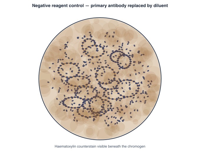
A CD20 immunohistochemistry run finishes and the technologist checks the controls. The negative reagent control section, shown here, carries diffuse brown chromogen across the tissue, and the patient section on the same slide rack is broadly brown as well. What does this tell the technologist about the run?
Show data table
| Run | Value (°C) | SD from mean |
|---|---|---|
| D1 | -25.2 | -0.2 |
| D2 | -24.6 | +0.4 |
| D3 | -25.4 | -0.4 |
| D4 | -25 | +0.0 |
| D5 | -24.8 | +0.2 |
| D6 | -25.3 | -0.3 |
| D7 | -24.9 | +0.1 |
| D8 | -25.1 | -0.1 |
| D9 | -23.5 | +1.5 |
| D10 | -23.4 | +1.6 |
| D11 | -23.6 | +1.4 |
| D12 | -23.3 | +1.7 |
| D13 | -23.5 | +1.5 |
| D14 | -23.4 | +1.6 |
Daily cryostat chamber temperature readings are plotted on the chart below, with the established mean at −25.0 °C and one standard deviation equal to 1.0 °C. The first eight days scatter closely around the mean; the last six sit together at roughly −23.5 °C without climbing further. How should this pattern be described?
Proving new reagents and equipment before patient use
Nothing new goes straight onto patient tissue. A fresh lot of eosin, a new antibody clone, a different paraffin, a replacement processor or a newly serviced microtome all have to demonstrate that they perform at least as well as what they replace. In practice this means staining control tissue of known behaviour side by side with the material in current use and comparing the sections under the microscope. Antibodies need tissues expected to stain and tissues expected not to. The comparison, the operator and the date are recorded, and only then does the item enter service.
A shipment arrives containing a new lot of alcoholic eosin from the usual supplier, and the routine haematoxylin and eosin stainer is due for a bath change the same morning. The technologist has control tissue available on the bench. What has to happen before patient sections meet the new lot?
A laboratory is bringing a newly purchased antibody clone into its immunohistochemistry menu and has completed the optimisation runs for dilution and retrieval. The technical specialist is preparing the verification package before any patient case is reported with it. What does that verification chiefly require?
Managing bench stock and reagent supply
Inventory work keeps the bench running without waste. It covers knowing what is on the shelf, ordering before stock runs out, receiving and checking deliveries against the order, recording lot numbers and expiry dates, and storing items as the supplier directs. Stock is rotated so the oldest usable container is drawn down first and newer stock sits behind it, which keeps material from quietly reaching expiry at the back of a cupboard. Expired reagents are removed from the working area rather than left available. Minimum stock levels for critical items are set so a routine order never becomes an emergency.
While restocking the special stains bench a technologist finds two bottles of periodic acid: one opened three weeks ago and still two-thirds full, and one unopened container delivered yesterday. Both carry expiry dates well into next year and both have been stored correctly. Which practice should be followed?
Handling case data and patient information
Histology generates identifiable data at every step: requisitions, accession numbers, block and slide labels, scanned images, and the reports themselves. The technologist is responsible for entering it accurately, keeping the link between patient, specimen, block and slide unbroken, and releasing it only through approved routes to people entitled to receive it. Whole-slide images and photographs count as patient records even when a name does not appear on the file. Personal email, removable drives and shared logins fall outside the laboratory's controlled paths, so requests that would use them are redirected rather than accommodated, however senior the requester.
A pathologist preparing a conference talk asks a technologist to send a scanned whole-slide image of a recent liver case to a personal email address, saying it will be quicker than going through the digital pathology system. How should the technologist respond?
Judging whether a stained section is fit to read
This is the histology-specific piece of quality work: looking at the finished slide and deciding whether a pathologist can safely read it. The technologist checks nuclear and cytoplasmic balance, differentiation, the colour a special stain has produced against its control, and whether processing or cutting has left a defect such as folds, chatter, knife lines or an out-of-focus scan. Faults affecting a whole rack usually point at a bath or reagent; faults on one slide point at the block or the section. An unacceptable slide is recut and restained rather than sent out with a caveat.
A full rack of routine sections comes off the automated stainer and every slide looks the same under the microscope: nuclei are faint and washed out while cytoplasmic and connective tissue staining is of normal intensity. Which explanation fits this finding?
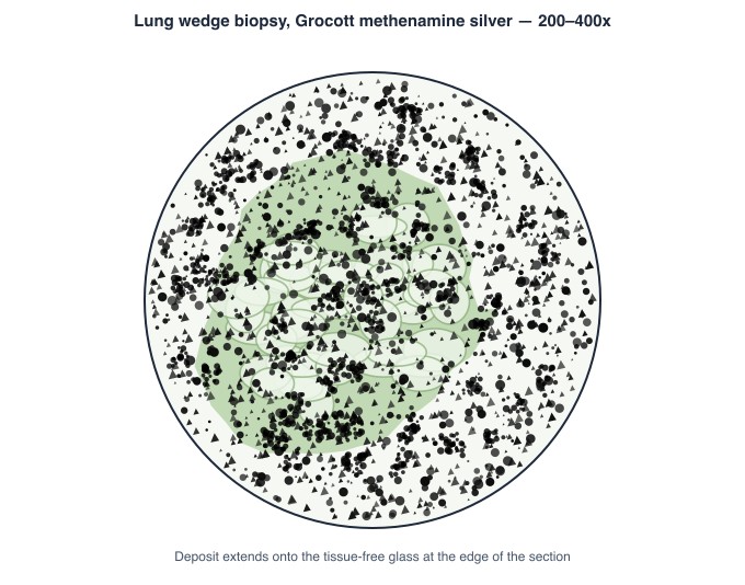
A Grocott methenamine silver on a lung biopsy is checked at the microscope before release. Dense black granular deposit lies over the whole section, as shown, obscuring tissue detail, and the fungal control stained in the same run is equally dark. What should the technologist do with it?
Working only from controlled procedures
Every method in the laboratory exists as a controlled document carrying a version number, an effective date and evidence of review. The technologist works from the current version only, and knows where to find it. Personal shortcuts, photocopied pages taped to cupboards and annotated printouts sit outside that system, and once a procedure is revised those copies quietly become wrong. Changes are made through the review process rather than by editing a bench copy. Obsolete versions are withdrawn from circulation, and staff sign to show they have read a revision before it takes effect.
Taped inside a cupboard door at the special stains bench is a photocopied page describing the Grocott method, and staff on that bench have clearly been using it. Comparing it with the current controlled version, a technologist sees the taped page gives a longer silver incubation. What should be done?
Keep reading with a free account
The rest of this page — 8 more sections and 12 practice questions — is free to read with an MLTPrep account.
No card required.
Back to all histology areas in the Histology study guide.
MLTPrep is an independent study resource. Not affiliated with or endorsed by CAMLPR. Always confirm current requirements with CAMLPR directly.