Histology
Preparation and Testing
Preparation and testing is the largest part of the histology exam because it is the largest part of the job: fixation, processing, embedding, microtomy, staining and microscopy, plus every judgement made along the way about whether the work is good enough to hand on. Almost none of it can be undone — an under-processed block, a torn section or an over-differentiated stain costs tissue that may be the only material a patient will ever give. The competency is as much about reading failures backwards to their cause as it is about running the method correctly the first time. Preparation and Testing is one of the eight competency areas assessed in the Histology Fields-of-Practice examination set by CAMLPR.
Making up and verifying bench reagents
Reagent work sits underneath every stain in the laboratory. A technologist weighs and dissolves the constituents, checks concentration and pH, filters what needs filtering, and labels the bottle with the preparation date, the expiry and the initials of whoever made it. Fixatives, decalcifying acids, differentiators and counterstains behave predictably only when made to formula, so calculations from concentrated stock have to be right the first time. New batches are checked against a section with known staining before they touch patient tissue, and a batch that fails is discarded rather than adjusted at the bench.
A technologist is making up one litre of 10 per cent neutral buffered formalin for the grossing bench, starting from a 37 to 40 per cent formaldehyde stock solution. What volume of stock solution is measured out?
A batch of Harris haematoxylin is being made up from raw constituents. Which component forms the coloured complex that anchors the dye to nuclear chromatin?
Bench methods run and judged by hand
Not everything in a histology laboratory runs on an instrument. Frozen sectioning, decalcification and its endpoint checks, manual coverslipping, macroscopic measurement and hand-run methods all depend on the technologist judging the procedure as it happens. The work calls for knowing why a method is done by hand at all — a frozen section exists precisely because paraffin processing would strip out the lipid or enzyme activity being looked for. Timing, temperature and reagent condition are recorded as the run proceeds, so a result can later be defended against the conditions that produced it.
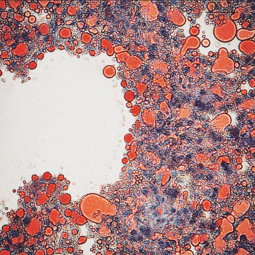
A liver biopsy from a patient investigated for a metabolic disorder is frozen, sectioned on the cryostat and stained with Oil Red O. The image shows the result. Why was this material not processed to paraffin in the usual way?
A bone trephine has spent 18 hours in 10 per cent formic acid. A sample of the used decalcifying fluid is drawn off and treated with ammonium hydroxide followed by ammonium oxalate, and no cloudiness develops. What does this indicate?
Running haematoxylin and eosin at the bench
Haematoxylin and eosin is the section a pathologist reads first, and on a manual bench its quality rests entirely on the technologist. You control the haematoxylin time, the depth of acid differentiation, the bluing step that fixes the nuclear colour, and the eosin that separates cytoplasm from background. Alcohols and xylene have to be clean and in the right sequence or the section clouds. Every rack is checked at the microscope before it leaves the bench, because a pale or over-differentiated batch is far cheaper to restain than to re-cut.
A manually stained batch of sections shows uniformly weak nuclear staining across the whole slide, while the eosin counterstain is of normal intensity. The haematoxylin was replaced with a fresh batch that morning. What is the most likely cause?
After the haematoxylin and the acid alcohol steps, sections are rinsed in a weakly alkaline solution such as Scott's tap water substitute. What does this rinse accomplish?
Setting up and using the microscope
A technologist looks down the microscope constantly — to check that a section is complete and cleanly stained, to confirm a special stain has worked before releasing the slide, and to screen preparations where that falls within scope. The instrument has to be set up properly: Koehler illumination, the correct objective, and the condenser and iris matched to the aperture in use. Some findings appear only under a particular configuration, so knowing when to reach for polarising filters, an oil immersion lens or a fluorescence cube is part of the competency.
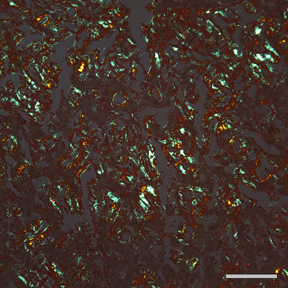
A skin biopsy stained with Congo red is being examined for amyloid, and the technologist obtains the appearance shown in the image. Which microscope configuration produces it?
Driving the tissue processor
Automation in histology means the tissue processor and the equipment around it. The technologist chooses a schedule to suit the specimens loaded, verifies reagent levels and rotation position, starts the run, and understands what the vacuum, pressure and heated stations each contribute. Blocks that reach the microtome soft, wet or brittle are read backwards to the schedule that produced them. Alarms, aborted runs and reagent exhaustion are handled and documented rather than restarted blindly, because under-processed blocks can usually be melted down and put back through the schedule, while tissue that has been over-processed and turned brittle cannot be brought back.
| Station | Reagent | Time |
|---|---|---|
| 1 | 10% neutral buffered formalin | 30 min |
| 2 | 10% neutral buffered formalin | 30 min |
| 3 | 70% ethanol | 20 min |
| 4 | 95% ethanol | 20 min |
| 5 | Absolute ethanol | 20 min |
| 6 | Absolute ethanol | 20 min |
| 7 | Xylene | 15 min |
| 8 | Xylene | 15 min |
| 9 | Paraffin wax, 60 °C | 30 min |
| 10 | Paraffin wax, 60 °C | 30 min |
The overnight schedule used on a routine automated processor is shown in the table. A 4 mm thick breast block run on this schedule sections badly, with a soft, wet centre that drags and will not ribbon. Which change to the schedule is indicated?
A vacuum-assisted enclosed tissue processor applies negative pressure during its paraffin stations. What does the vacuum contribute at that point in the run?
Selecting and controlling special stains
Special stains answer questions the routine section cannot: is this collagen or muscle, is fungus present, is the mucin acidic or neutral, is there amyloid. The technologist selects the method for the question asked, runs a known positive control alongside the patient section, and reads the control first. Many of these methods hinge on a single controllable variable — a pH, a differentiation time, a silver impregnation endpoint watched under the microscope. Reagent age and section thickness matter more here than in routine work, and a failed control invalidates the whole run.
Two sections of a gastric biopsy are stained with alcian blue, one buffered to pH 2.5 and one to pH 1.0. Material that stains blue on both sections has which composition?
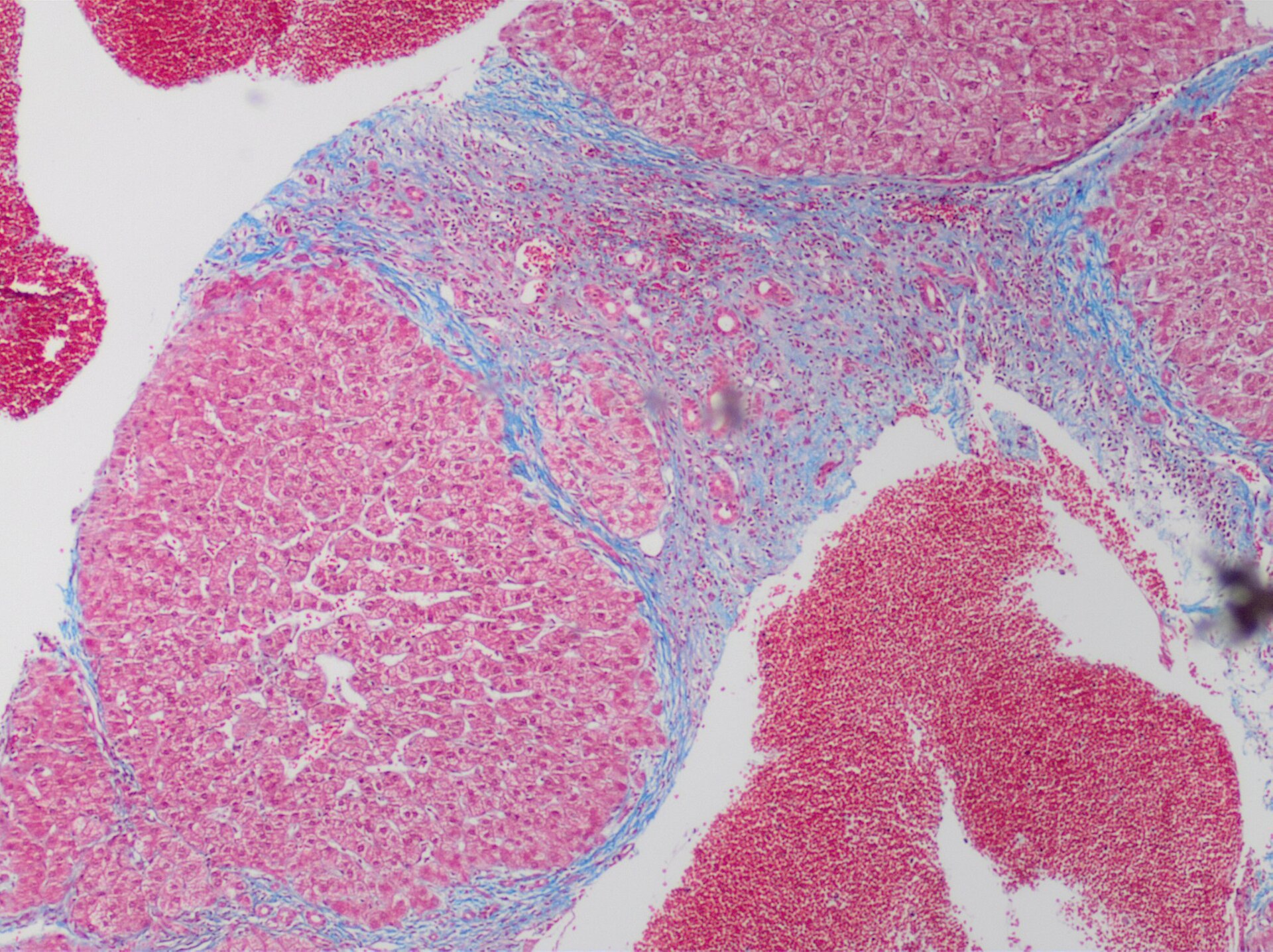
A liver biopsy stained by Masson's trichrome shows the blue-green bridging bands seen in the image. Which step in the trichrome sequence governs whether collagen retains that blue-green dye?
Grossing, embedding and cutting sections of tissue
This is the craft at the centre of the discipline. It starts at the grossing bench, where the specimen is described, measured and sampled into cassettes so the right tissue reaches the block, and continues with turning a processed block into a flat, complete, correctly oriented section on glass. Embedding sets the orientation the pathologist will see. Trimming, block cooling, knife angle, section thickness, flotation bath temperature and drying all have to be right together, and each has its own failure signature — chatter, knife lines, folds, holes, thick and thin banding. A technologist reads those signatures at the microscope and corrects the cause rather than cutting more sections and hoping. Levels, serials and control sections are cut to whatever protocol has been requested.
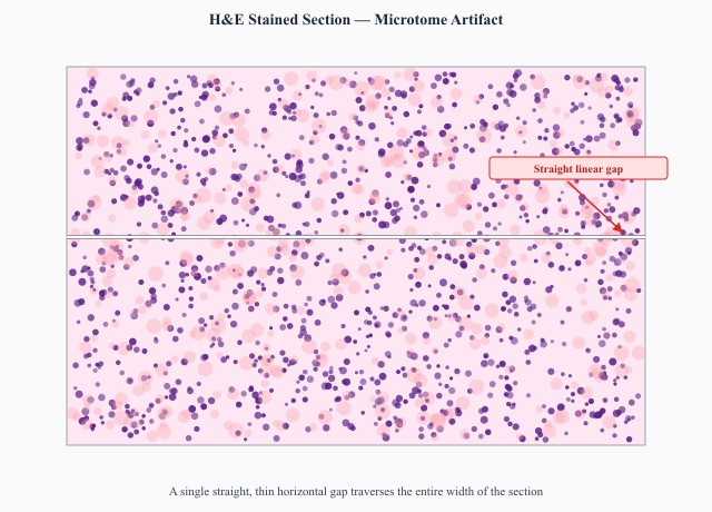
Sections cut from a routine paraffin block carry a thin straight line running parallel to the cutting direction, as shown in the image, and it appears on every section in the ribbon at the same position. What is the immediate corrective action?
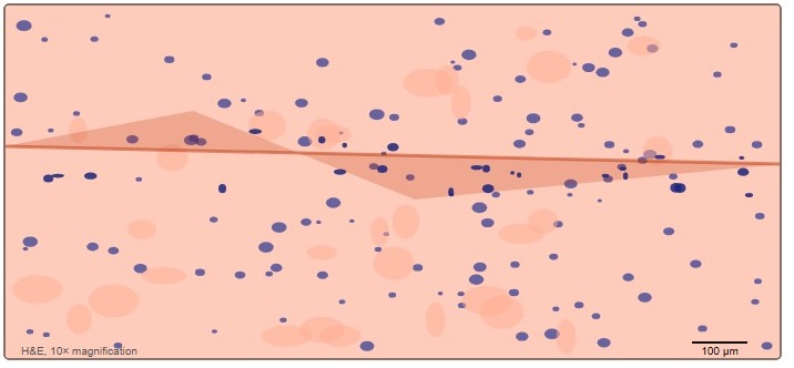
Sections lifted onto slides show doubled tissue layers and persistent surface folds, as in the image. The flotation bath is reading 42 °C and the blocks are embedded in a wax with a melting point of 58 °C. What should be adjusted?
Preparing smears, imprints and cytospins
Some preparations never see a paraffin block. Cytology smears, fine-needle aspirates, touch imprints and cytospins are made directly from cells and behave quite differently from a cut section. The critical decision is usually fixation: material intended for Papanicolaou staining must be fixed while the slide is still moist, whereas Romanowsky methods want it air-dried. Spreading pressure, cell density and slide labelling all determine what can be read afterwards, and preparations made away from the laboratory need the same identity and fixation discipline as those made at the bench.
A cervical smear is spread onto a slide at the clinic, then travels to the laboratory for Papanicolaou staining. At what point must the slide be fixed?
Judging whether a specimen can give a valid result
Before any work starts, the technologist decides whether the specimen can support a valid result. That means checking identity against the requisition, the container, the fixative used, the ratio of fixative to tissue, the thickness of the slices and how long the specimen sat before it reached formalin. Under-fixed, dried, crushed or frozen-and-thawed tissue produces changes that survive into the section and can be mistaken for pathology. Where a defect can be corrected — re-slicing a thick piece, topping up fixative — it is corrected and recorded; where it cannot, the limitation is passed to the pathologist.
A bone marrow trephine section shows streamed, elongated nuclei and smeared chromatin across much of the core, while a small area at one end is well preserved. Which pre-analytical event best explains the appearance?
A uterine leiomyoma arrives in the laboratory as a single slab about 25 mm thick, sitting in roughly 30 mL of formalin in a small container. What should be done before the specimen goes onto a processor?
Testing performed at the patient's side
Point-of-care work takes the laboratory out to the patient. In histology that mostly means supporting intra-operative and on-site services: preparing and rapidly staining aspirate smears or touch preparations at the bedside so adequacy can be judged before the needle is withdrawn, and running frozen sections beside the operating theatre. The same rules still apply away from the bench — controls run and recorded, operators trained and signed off, instruments maintained, results entered into the permanent record. Speed is the whole point of the service, but it never displaces identification or quality control.
A technologist supports rapid on-site evaluation in the endoscopy suite, preparing air-dried smears from each fine-needle pass and staining them at the bedside so the operator can be told within a minute whether the sample is adequate. Which stain is used for this?
Running and checking automated stainers
Automated stainers carry most of the routine haematoxylin and eosin workload and nearly all of the immunohistochemistry. The technologist loads racks, confirms programme assignment, keeps reagents topped up and rotated on schedule, and reviews the output rather than trusting it. Immunohistochemistry runs carry their own controls — a positive control section for each antibody and a negative reagent control — and both are read before any patient slide is scored. Drift in colour intensity across a day usually points at reagent carryover or exhaustion, and instrument maintenance is logged as part of the record.
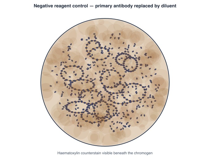
On an automated immunohistochemistry run, the negative reagent control section shows the diffuse brown chromogen seen in the image. The positive control on the same run stains as expected. What happens to the patient slides from that run?
An automated linear haematoxylin and eosin stainer runs 300 slides over a shift. Racks stained early in the day look normal, but the eosin becomes progressively paler through the afternoon and the last racks are noticeably weak. Which routine practice prevents this?
Keep reading with a free account
The rest of this page — 10 more sections and 17 practice questions — is free to read with an MLTPrep account.
No card required.
Back to all histology areas in the Histology study guide.
MLTPrep is an independent study resource. Not affiliated with or endorsed by CAMLPR. Always confirm current requirements with CAMLPR directly.