Histology
Equipment and Resources
Histology depends on instruments that have to be right before the first block is cut: microtomes, tissue processors, flotation baths, embedding centres, cryostats, stainers, coverslippers and slide scanners. This domain covers running them inside specification, keeping the maintenance and calibration record that proves it, reading the signature of a failing instrument in the sections it produces, and capturing digital images that reproduce faithfully what sits on the glass. Equipment and Resources is one of the eight competency areas assessed in the Histology Fields-of-Practice examination set by CAMLPR.
Keeping instruments running, verified and documented
Histology equipment rarely fails all at once; it drifts. This area covers the daily, weekly and monthly work that holds a microtome, tissue processor, flotation bath, embedding centre, cryostat and stainer inside their working range: checking and recording bath and paraffin temperatures, confirming cryostat chamber temperature before the first frozen section, rotating and topping up processor reagents on schedule, cleaning and lubricating the microtome, and logging every check so slow drift becomes visible. The expectation is not simply to operate the instrument but to confirm it is performing and to record that confirmation.
A technologist charges the flotation bath before cutting a tray of blocks whose embedding medium melts at 56 degrees Celsius. At what setting will ribbons flatten properly without the sections coming apart?
| Station | Reagent | Blocks since last change |
|---|---|---|
| 1 | 70% ethanol | 640 |
| 2 | 95% ethanol | 640 |
| 3 | 95% ethanol | 640 |
| 4 | Absolute ethanol | 640 |
| 5 | Absolute ethanol | 640 |
| 6 | Absolute ethanol | 640 |
The dehydration reagents on an enclosed tissue processor are scheduled for rotation, and the log for the alcohol stations is shown [TABLE]. Tissue passes through the stations in numerical order. When the rotation is carried out, which station receives the fresh bottle of absolute alcohol?
Recognising when the instrument is the fault
When section quality falls away, the technologist has to work out whether the fault sits in the tissue, the reagents or the instrument, and then act. This area covers reading a defect back to its source: a line repeating in the same place pointing to a damaged knife edge, alternating thick and thin sections pointing to a loose clamp, ribbons that will not form pointing to clearance angle or a cold block face, frost inside a cryostat pointing to a door left ajar. It also covers the limits of bench correction, and knowing when to withdraw an instrument, tag it and call service.
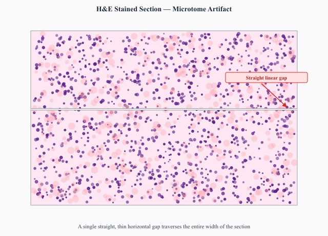
Every section from one paraffin block carries the same fine straight line running parallel to the direction of cutting [IMAGE]. The block is refaced and the line returns in exactly the same position. What should the technologist do next?
Ribbons from a well-fixed uterine block come off a rotary microtome alternating thick and thin within the same ribbon, and a second block cut immediately afterwards behaves the same way. Which part of the setup should be checked first?
Digital slide capture and image quality
Digital capture is now routine histology work: photomicrographs for reports and teaching files, gross specimen photographs at the cut-up bench, and whole-slide scanning for consultation and archiving. This area covers operating the camera or scanner properly — balancing colour against a clear field so stains reproduce truly, matching magnification and resolution to the purpose, laying enough focus points across a section that uneven or thick areas stay sharp, including a scale bar where size matters, and keeping every file tied to the correct accession number. A technically sound slide is worthless if the captured image misrepresents it, so images are reviewed before release.
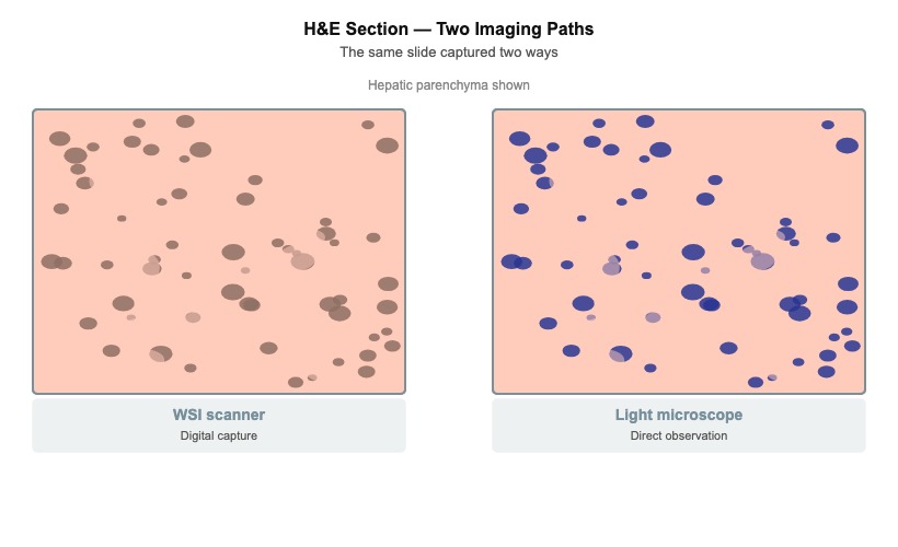
Slides scanned overnight are opened the next morning and every haematoxylin and eosin image in the run looks warm, with nuclei reproducing brownish-pink instead of blue-purple [IMAGE]. Which scanner step will correct the run?
A large bowel resection section is scanned for a case conference. On review the centre of the tissue is soft and blurred while the peripheral tissue is crisp. What is the appropriate next step?
Keep reading with a free account
The rest of this page — 2 more sections and 4 practice questions — is free to read with an MLTPrep account.
No card required.
Back to all histology areas in the Histology study guide.
MLTPrep is an independent study resource. Not affiliated with or endorsed by CAMLPR. Always confirm current requirements with CAMLPR directly.