Hematology
Preparation and Testing
Preparation and testing covers everything a technologist does between accepting a specimen and generating a result someone can act on: confirming identity at each handover, preparing reagents and controls, spreading and staining films, driving the analyser, and reading what is actually on the glass. Much of the work is manual craft — a badly spread film or an over-buffered stain destroys morphology no instrument can recover. It also covers knowing where each method stops being reliable, because interference that goes unrecognised becomes a released result. Preparation and Testing is one of the eight competency areas assessed in the Hematology Fields-of-Practice examination set by CAMLPR.
Keeping the tube and the slide tied to one patient
Identity has to survive every step between the collection chair and the report. On receipt you check that the name and unique number on the tube agree with the requisition, and you check again whenever the specimen changes container or moves onto glass. Films, cytospins and marrow preparations carry their own written identity in pencil, because a slide separated from its tube proves nothing about whose blood it holds. Where the tube and the paperwork disagree, you do not correct the label: the specimen is held, the discrepancy documented, and a fresh collection requested.
Four wedge films are spread from a single EDTA tube for a haematology consultation, then carried to the staining bench with several other batches. How should the films be identified before they leave the spreading bench?
Bringing reagents, calibrators and controls into a usable state
Reagents, calibrators, standards and controls behave as the manufacturer intended only if they are prepared exactly as the insert describes. That means reconstituting with the stated diluent and volume, allowing full dissolution, mixing by gentle inversion rather than shaking, and bringing material to its working temperature before sampling. Every vial is dated when opened and discarded at whichever comes first, the printed expiry or the on-board stability. Lot numbers, preparation dates and the technologist's initials are recorded, so that a control shift noticed weeks later can be traced back to the material behind it.
A lyophilised APTT control is reconstituted with the exact volume of diluent named in the package insert. Before any of it is drawn into the analyser, what should the technologist do with the vial?
A bottle of lysing reagent printed with a manufacturer expiry of 30 November is opened on 1 September. The insert states the reagent is stable for 30 days once opened. What expiry should be written on the bottle?
Staining films so the morphology can actually be read
A Romanowsky stain balances an acidic eosin against a basic azure dye, and the buffer decides which one wins. Too alkaline and the whole film turns blue-grey with inky nuclei; too acidic and nuclei fade while red cells go brick orange. You control buffer pH, fixation in absolute methanol, staining and rinse times, and you examine a stained film before releasing a batch. Some findings need a supravital stain instead — new methylene blue for reticulocytes, crystal violet for Heinz bodies — because those inclusions are simply not there on a fixed, methanol-treated preparation.
Every film from the morning batch is uniformly too blue: red cells look grey-blue, nuclei are inky and dense, and cytoplasmic detail in the leucocytes is lost. The buffer in use measures pH 7.4 against a procedural requirement of 6.8. What corrects the batch?
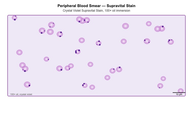
The preparation in the image was made by incubating red cells with crystal violet and examining the film without any fixation. What does this technique reveal that a routine Wright-Giemsa film cannot?
Naming every element in the field
This is the identification work underpinning every differential. You recognise each granulocyte stage from promyelocyte through band form, separate a reactive lymphocyte from a blast, tell an eosinophil from a neutrophil carrying toxic granulation, and pick out red cell inclusions and platelet abnormalities. Judgements rest on repeatable criteria — nuclear shape and chromatin texture, presence of a nucleolus, granule size and colour, the nuclear to cytoplasmic ratio — rather than on impression. Cells you cannot name are referred to a senior technologist or a pathologist rather than guessed at, because one misidentified blast redirects a whole investigation.
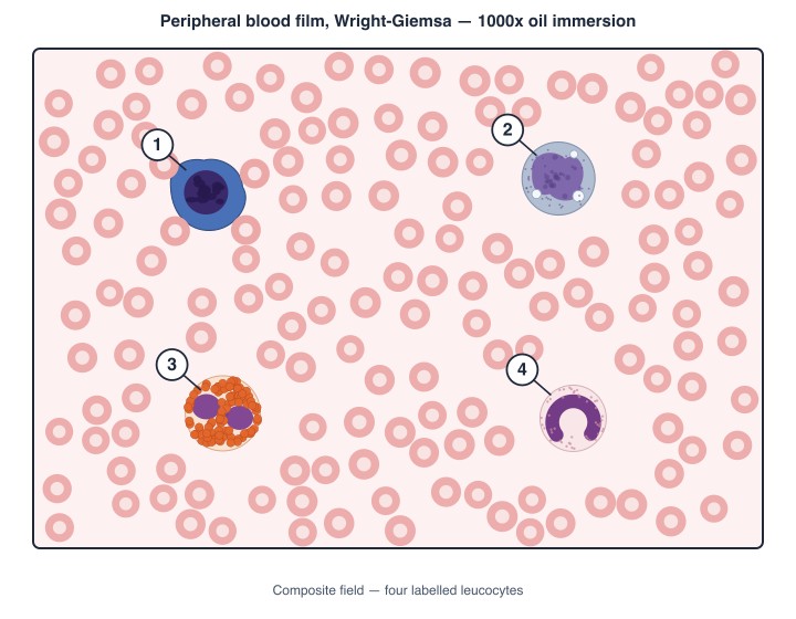
In the composite field shown, cell 1 is large, its plentiful dark blue cytoplasm flattening against every red cell it touches, with coarsely clumped chromatin and no nucleolus in view. How should it be recorded on the differential?
Which granulocytic precursor is the most mature stage still able to divide?
Knowing where the analyser stops being right
Every method has a range in which it can be trusted and conditions that push it outside. An impedance counter cannot tell a platelet clump from a red cell, reads a lysis-resistant nucleated red cell as a leucocyte, and takes turbidity from lipid or a very high white count as haemoglobin. Cold agglutinins collapse the red cell count and inflate the mean cell volume. Recognising the pattern — an impossible MCHC, a flagged histogram, indices that contradict the film — and knowing the corrective procedure is what separates a released result from a wrong one.
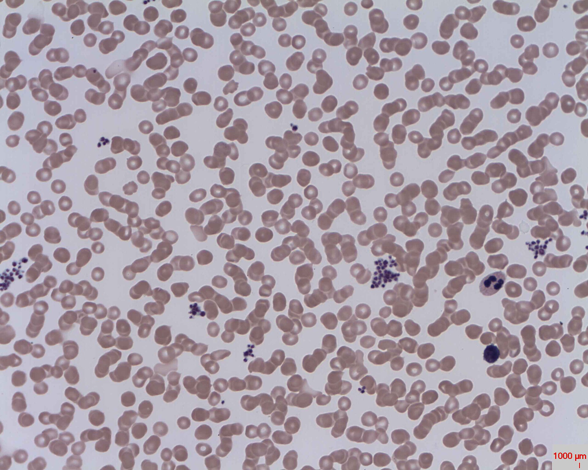
A specimen sent in from a community clinic triggers a platelet-clump flag and reports 38 ×10⁹/L. The tube holds no visible clot, and film review shows the appearance in the image. What will give a usable platelet count?
A specimen from a patient with untreated chronic lymphocytic leukaemia gives WBC 310 ×10⁹/L, haemoglobin 121 g/L, haematocrit 0.22 L/L and a calculated MCHC of 550 g/L. Which explanation fits this set of numbers?
Finding parasites and other organisms on the film
A blood film is a diagnostic specimen for organisms as well as for cells. Thick films concentrate a large volume of blood and find low-level parasitaemia; thin films keep red cell size and shape intact so a species can be named and the percentage of infected cells counted. You look for ring forms, gametocytes, malarial pigment, stippling and the size of infected cells, and also for organisms inside leucocytes, microfilariae and trypanosomes swept to the feathered edge. A positive finding is urgent, and negative films never close the question — serial collections are the standard.
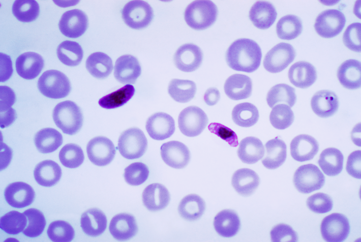
A thin film from an agricultural worker who spent six weeks in West Africa shows the appearance in the image: normal-sized red cells, a delicate ring form carrying two chromatin dots, and crescent-shaped gametocytes with a band of central pigment. Which infection is present?
Both a thick and a thin film are prepared on every specimen submitted for malaria investigation. What does the thick film add that the thin film cannot supply?
Making a film that is worth reading
A film that is too thick, too short or streaked cannot be rescued by anything the stain does afterwards. Spreading angle, drop size and stroke speed are matched to the specimen: a thick, polycythaemic sample needs a shallower angle and a smaller drop, an anaemic one the opposite. Films are air-dried quickly and completely, never breathed on or heated, since trapped moisture leaves refractile artefact through the red cells that mimics inclusions. Body fluids with few cells go through a cytocentrifuge instead, concentrating the cells into a small readable circle without tearing them.
Films spread from a specimen with a haematocrit of 0.68 L/L are short, thick and dark, and no monolayer forms anywhere along their length. What adjustment should be made when the films are respread?
A cerebrospinal fluid with 4 ×10⁶/L nucleated cells is received with a request for a differential count. Which preparation will put enough intact cells in one place to allow the count?
Setting up a molecular assay without contaminating it
Molecular work in a haematology laboratory — JAK2, BCR-ABL1, factor V Leiden — depends on set-up discipline far more than on the thermal cycler. Reagent preparation, template addition and amplification occupy physically separate areas, and staff, gowns, pipettes and racks move one way only, so amplified product can never reach a clean reaction mixture. Nucleic acid is extracted from EDTA or citrate blood and never from heparin, which inhibits the polymerase. Every run carries positive, negative and no-template controls, and a no-template control that amplifies invalidates the run rather than prompting a report.
A laboratory introducing a JAK2 V617F assay assigns reagent preparation, specimen addition and amplification to three separate rooms, and requires staff, coats and pipettes to travel in one direction only through them. What does this arrangement protect against?
Deciding whether the specimen can be tested at all
Before anything is analysed, the specimen has to be capable of giving a valid answer. You check that the tube type suits the test, that it is filled to the mark, that it was mixed and is free of clots, and that its age and storage still fall inside the acceptable window. Coagulation is the least forgiving: an underfilled citrate tube leaves excess anticoagulant relative to plasma. A clot in an EDTA tube consumes platelets and leucocytes as it forms, so the count is wrong even though the analyser reports it perfectly happily.
A 2.7 mL sodium citrate tube reaches the coagulation bench filled to roughly 1.8 mL, with a request for PT and APTT. If it were tested as received, what would happen to the results?
Checking an EDTA tube with wooden applicator sticks reveals a small dark clot clinging to one stick. The analyser has already generated a complete result set on that tube. What should be done with it?
Reading the cells and everything else in the field
Microscopy is not only about naming cells. You scan at low power for cell distribution, rouleaux, agglutination, platelet clumps, fibrin strands and stain deposit before dropping to oil for detail. Non-cellular findings carry as much information as the cells themselves: a blue-tinged background with red cells stacked in columns points toward a paraprotein, fibrin strands say the tube clotted, and refractile spots say the film dried badly. In body fluids the same discipline covers crystals, casts and debris. Recording what is there, and separating a real finding from an artefact, is the whole task.
A film from a 68-year-old investigated for back pain and fatigue shows red cells stacked in long columns like coins, with the plasma spaces between them staining diffusely blue. Which follow-up investigation does this pattern most directly point toward?
Scanning a stained film at low power, a technologist finds pale pink strands running in parallel across one region of the film, with leucocytes and platelets caught along them and stripped from the surrounding area. What do the strands represent?
Separating the components of a specimen and measuring them
Some answers only appear once the parts of a specimen are pulled apart. Haemoglobin variants are separated by high performance liquid chromatography, capillary electrophoresis or gel, and each fraction is quantitated: a raised HbA2 with mild microcytosis and normal iron studies is a very different result from an isolated abnormal band. Qualitative screens such as the solubility test say only that a sickling haemoglobin is somewhere in the sample, so a separation method must always follow before anything is named. Plasma for coagulation is separated by centrifugation, with the fractions handled to preserve the analyte.
| Fraction | Result (%) | Reference (%) |
|---|---|---|
| HbA | 92.0 | 96.0 - 98.0 |
| HbA2 | 5.4 | 2.0 - 3.5 |
| HbF | 2.6 | less than 1.0 |
| Abnormal band | none detected | none |
The chart gives haemoglobin fractions measured by high performance liquid chromatography on a 4-year-old with a mild microcytic picture, a normal ferritin and a normal serum iron. Which interpretation fits the fractions shown?
A haemoglobin solubility screen run on a 22-year-old attending a pre-operative clinic gives a turbid, opaque tube through which the printed lines cannot be read. What must be done before any result is issued?
Putting a number on what is under the objective
Counting is what turns morphology into something a clinician can act on. Manual differentials run over a set number of cells in the monolayer using a systematic pattern, and nucleated red cells are tallied separately so the white count can be corrected for them. Platelet estimates average several oil fields and multiply by a factor specific to that microscope, then get checked against the analyser. Chamber counts, reticulocyte percentages and absolute counts follow defined criteria and replicate limits. Semi-quantitative grading of features such as polychromasia uses the laboratory's published scale, not personal judgement.
Working in the monolayer with a 100× oil objective whose validated field factor is 20, a technologist counts platelets across ten consecutive fields and averages 12 per field. What is the estimated platelet count?
An analyser reports a white cell count of 22.5 ×10⁹/L on a newborn. The 100-cell differential performed on the film includes 25 nucleated red cells seen per 100 leucocytes. What white cell count should be reported?
Producing a complete blood count you can stand behind
The complete blood count is the workhorse of the department, and running it well means much more than pressing start. You confirm the specimen is adequate, review every flag and histogram, and check that the indices hang together internally: haemoglobin roughly a third of the haematocrit, an MCHC inside plausible limits, the rule of three holding. Results failing those checks are investigated, not reported. Films are made wherever the review criteria demand one, critical values are telephoned immediately, and delta checks against previous results are resolved before release rather than after a clinician calls.
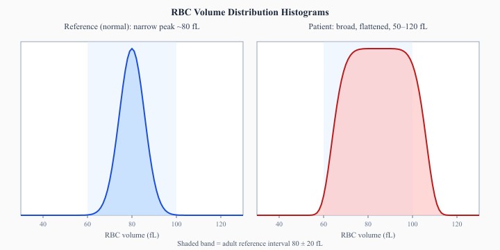
The image compares a patient's red cell volume distribution against a reference tracing. The patient curve is broad and flattened, spreading from roughly 50 fL to 120 fL, where the reference curve is a narrow peak near 80 fL. Which reported index will be raised?
A count on a well outpatient attending for a routine renewal returns a haemoglobin of 96 g/L with a haematocrit of 0.42 L/L. There are no analyser flags. Applying the rule of three, what should the technologist do?
Testing plasma that can carry a coagulation result
Coagulation testing measures a reaction happening in plasma, so nearly everything that goes wrong has already gone wrong before the clot forms. Blood is drawn into 3.2 per cent sodium citrate at the correct ratio, centrifuged to strip out platelets, and tested inside the stability window for that assay. Screening tests — PT with INR, APTT, fibrinogen and thrombin time — are followed by mixing studies, factor assays and inhibitor work when they come back prolonged. Controls at two levels run with every batch, and a shift in the control pattern stops reporting until it is explained.
Show data table
| Run | Value | SD from mean |
|---|---|---|
| 1 | 30.5 | -0.4 |
| 2 | 30.6 | -0.3 |
| 3 | 31.4 | +0.3 |
| 4 | 31.6 | +0.5 |
| 5 | 31.3 | +0.3 |
| 6 | 31.8 | +0.7 |
| 7 | 31.5 | +0.4 |
| 8 | 31.9 | +0.8 |
| 9 | 31.6 | +0.5 |
| 10 | 32.0 | +0.8 |
| 11 | 31.7 | +0.6 |
| 12 | 32.1 | +0.9 |
The chart plots the normal-level APTT control across twelve consecutive runs, with an established mean of 31.0 s and an SD of 1.2 s. No single point falls outside 2 SD. Which control rule has nonetheless been violated?
Citrated specimens collected for a lupus anticoagulant panel are to be frozen at −70 °C and shipped to a reference laboratory. Which requirement must the separated plasma meet before it goes into the freezer?
Catching haemolysis, lipaemia and icterus before they reach a result
Haemolysis, lipaemia and icterus alter results by real mechanisms, not simply by looking unpleasant. Free haemoglobin from a traumatic draw stays in the measuring cuvette while the red cells that carried it are gone, so the haematocrit falls, the haemoglobin holds, and the MCHC climbs past any believable value. Lipid turbidity is read by the photometer as haemoglobin and raises it falsely. You inspect the plasma layer at every stage, grade what you see against the laboratory's scale, and apply the stated remedy — a fresh collection, or a plasma replacement step — commenting on any interference that remains.
A companion tube spun down beside a count shows a deeply red supernatant, and the collector notes the draw was slow and difficult through a 25-gauge needle. The count gives haemoglobin 118 g/L, haematocrit 0.24 L/L and MCHC 492 g/L. What is the correct action?
A specimen from a patient receiving a parenteral lipid infusion separates with a milky plasma layer. The count gives haemoglobin 158 g/L, haematocrit 0.31 L/L and MCHC 510 g/L, and the film shows normal red cell staining. Which procedure will give a usable haemoglobin?
Keep reading with a free account
The rest of this page — 14 more sections and 27 practice questions — is free to read with an MLTPrep account.
No card required.
Back to all hematology areas in the Hematology study guide.
MLTPrep is an independent study resource. Not affiliated with or endorsed by CAMLPR. Always confirm current requirements with CAMLPR directly.