Hematology
Evaluation and Interpretation
Evaluation and interpretation is where numbers and morphology turn into a decision. This domain covers judging whether a result is even possible, whether an abnormality belongs to the patient or to the specimen, what a set of haematology results means when read together, and when a finding must be confirmed, corrected or escalated before it leaves the laboratory. It is the judgement layer that sits between a validated instrument and a released report. Evaluation and Interpretation is one of the eight competency areas assessed in the Hematology Fields-of-Practice examination set by CAMLPR.
Sorting findings that matter from findings that do not
Not every abnormality on a film or an analyser printout changes what happens to the patient. This area asks you to separate findings that alter diagnosis, treatment or urgency from those that are incidental, age-appropriate or purely cosmetic. A single blast, a marked left shift, or a platelet count of 8 ×10⁹/L demands action; a few crenated cells near the feathered edge does not. You are expected to know your laboratory's film review criteria, apply them the same way on every shift, and escalate the findings that meet them rather than reporting everything you see with equal weight.
A 34-year-old attends for a pre-employment CBC. Every numerical result falls inside the reference interval, but film review turns up several incidental features. Which finding most clearly requires escalation to a pathologist?
A regular blood donor's screening film shows most neutrophils with only two nuclear lobes joined by a thin filament, with normal granulation and no abnormality in any other lineage. All counts are normal, and films from the donor's father show the same picture. What does this represent?
Telling a real abnormality from a specimen or analyser artefact
Analysers and stained films both produce appearances that are not real patient findings. Cold agglutinins, platelet satellitism, platelet clumping, drying and stain deposit, underfilled tubes, clotted specimens and delayed transport all imitate disease. The skill here is recognising the pattern, proving it — usually by inspecting the film, checking the specimen and its indices, or repeating under altered conditions such as warming to 37 °C — and then correcting or recollecting before a spurious number reaches the chart. Releasing an artefact as a result is a patient-safety event, not a technicality.
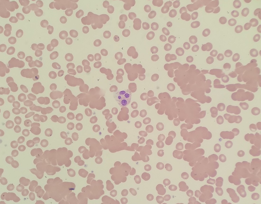
A CBC from a 71-year-old reaches the laboratory from the emergency department in January. The analyser reports RBC 1.9 ×10¹²/L, MCV 121 fL and MCHC 448 g/L, and the film shows the appearance in the image. What is the appropriate next step?
An analyser reports a platelet count of 41 ×10⁹/L on an outpatient who has no bruising or bleeding. There is no clot in the tube, and the film shows platelets rosetting around the neutrophils. What should the technologist do?
Reading blood and body fluid results as a whole
Results are read as a set, not one number at a time. You weigh the counts, indices, differential and film against reference intervals appropriate to the patient's age, and you check internal consistency — does the haemoglobin fit the haematocrit, does the MCV fit the morphology, does the RBC count fit the MCV. Body fluids carry their own rules: corrected counts after a bloody tap, cell-type expectations for CSF, synovial and serous fluids, and dilution decisions for viscous or heavily cellular samples. The interpretation you reach directs the next test and the urgency of the call.
A 26-year-old has Hb 112 g/L, RBC 6.1 ×10¹²/L, MCV 66 fL, RDW 13.0% and a ferritin within the reference interval. Red cells on the film are uniformly small, with occasional target cells. Which condition do these results most support?
CSF obtained from a traumatic lumbar puncture contains RBC 5000 ×10⁶/L and WBC 12 ×10⁶/L. Blood drawn at the same time shows RBC 4.0 ×10¹²/L and WBC 8.0 ×10⁹/L. How should the CSF white cell count be interpreted?
Verifying what a digital imaging system shows you
Digital cell-imaging systems present captured fields together with a suggested classification, not a finished differential. Your task is to confirm or reclassify every cell the system has pre-sorted, recognise the categories it cannot resolve reliably, and judge when the image itself is the problem — a field taken from too thick or too thin an area, poor focus, a stain that has drifted in colour, or overlapping cells read as one. The same discipline applies to images sent for remote review: decide whether the field is representative before drawing any conclusion, and go back to the glass slide when it is not.
A digital morphology system captures a red cell field and pre-classifies the cells as fragmented. The patient is 29 years old with platelets of 22 ×10⁹/L and a rising creatinine. What must the technologist do before the differential is released?
A rural site uploads digital blood film images for review at a referral centre. Every captured field was taken from the extreme feathered edge of the smear. What effect will this have on the differential?
Catching results that cannot be true
Some results are not merely abnormal — they are impossible, or impossible for this patient today. A haematocrit that does not match the haemoglobin, a platelet count that contradicts the film, a haemoglobin that has halved overnight with no bleeding: each points to a pre-analytical or analytical problem rather than to a change in the patient. You are expected to notice these before release, run the checks that identify the cause — delta comparison, film review, specimen inspection, repeat on a fresh draw — and document what you found and what you did about it.
A 3-year-old with severe iron deficiency has an MCV of 55 fL and an analyser platelet count of 780 ×10⁹/L. The film shows marked microcytosis with some red cell fragments, and platelets that look adequate but not increased. What is the most likely explanation for the platelet count?
A postoperative patient's haemoglobin was 128 g/L at 08:00 and is 74 g/L on a specimen received at 20:00 from the same ward. The patient is haemodynamically stable, the dressing is dry, and a repeat on the same tube gives an identical result. What should be suspected first?
Judging kit-based and point-of-care results
Kit-based tests still have to be judged, not simply read off. Before a result is released you confirm that the kit and its reagents are in date and were stored correctly, that the lot's controls and any built-in procedural control performed as expected, and that the reaction was read inside the stated time window. You also need each kit's limitations: what makes a sickle solubility screen falsely positive or falsely negative, why a positive screen still needs confirmatory testing, and which point-of-care results must be repeated on a laboratory analyser before they guide treatment.
A solubility screening test performed on an 18-year-old gives a turbid suspension that obscures the printed lines viewed through the tube. Kit controls performed as expected. What is required before a result is reported?
A point-of-care D-dimer cartridge returns a numerical result, but the device's built-in procedural control did not develop. What should be done with the result?
Keep reading with a free account
The rest of this page — 5 more sections and 10 practice questions — is free to read with an MLTPrep account.
No card required.
Back to all hematology areas in the Hematology study guide.
MLTPrep is an independent study resource. Not affiliated with or endorsed by CAMLPR. Always confirm current requirements with CAMLPR directly.