Clinical Microbiology
Preparation and Testing
Preparation and testing is where the microbiology bench does most of its work: turning a received specimen into a stained smear, an inoculated plate, a nucleic acid extract or a susceptibility panel. This domain covers identity checks at the bench, reagent and media preparation, smear and culture technique, microscopy, molecular set-up and antimicrobial susceptibility testing. Mistakes made here are rarely recoverable later, because the specimen is often consumed by the test. Preparation and Testing is one of the eight competency areas assessed in the Clinical Microbiology Fields-of-Practice examination set by CAMLPR.
Confirming the specimen in front of you belongs to the named patient
Before a single plate is inoculated, the technologist confirms that the container, the requisition and the laboratory record describe the same person. In microbiology this means at least two independent identifiers plus the collection site, date and time, because one patient may have several swabs taken from different sites on the same day. Missing labels, labels applied to the transport bag rather than the container, and site mismatches are resolved with the collecting area before processing. Specimens such as cerebrospinal fluid or intra-operative tissue cannot be recollected on demand, so identity problems are corrected with the requester rather than settled by rejection.
A wound swab arrives with the patient's full name and health-record number on the transport tube, but the requisition names a different collection site than the tube label. What should the technologist do first?
A cerebrospinal fluid specimen collected in theatre arrives labelled with the patient's surname only, and laboratory policy requires two identifiers. What is the appropriate action?
Preparing and proving reagents, media and control materials
Microbiology depends on reagents and media that behave predictably: Gram stain solutions, oxidase and catalase reagents, turbidity standards, broth diluents and prepared plates. The technologist reconstitutes materials exactly to the manufacturer's instruction, records lot numbers and expiry dates, and stores each item at the stated temperature and atmosphere. New lots are checked against organisms with known reactions before any patient use, and stock control organisms are subcultured on a set schedule so they stay viable and unchanged. Media that are dehydrated, haemolysed, contaminated or discoloured are removed from the bench rather than used cautiously.
A new lot of Gram stain reagents is opened at the start of a shift. Which check confirms the lot performs before patient smears are reported?
A 0.5 McFarland standard is needed to prepare an inoculum for disk diffusion. The tube has been standing on the bench and the contents have visibly settled. What should be done before it is used?
Staining smears so morphology and reaction are readable
Staining converts an invisible smear into information. The technologist fixes the smear appropriately, then applies the stain sequence with timings that suit the specimen. Decolourisation is the step most often mishandled: too long strips crystal violet from Gram-positive organisms, too short leaves Gram-negative organisms falsely purple. Acid-fast, modified acid-fast, calcofluor, India ink and lactophenol preparations each answer a different question, and the choice follows what the requester has asked. Every stained batch is read against a control slide before any patient interpretation is made, because a reagent fault affects the whole batch equally.

A stool specimen from a patient with three weeks of watery diarrhoea after a petting-farm visit is stained as shown. Which staining property makes these 4-6 µm structures appear pink against the background?
A batch of Gram stains prepared from pure Staphylococcus aureus cultures all read pink-red, and the control slide stained in the same batch looks the same. Which step should be reviewed first?
Recognising the microscopic elements that matter in a specimen
Beyond bacteria, the microbiology technologist recognises the other elements on a slide: neutrophils, squamous and other epithelial cells, yeast, hyphae, protozoal trophozoites and cysts, helminth ova and background debris. Each element changes how the culture is handled. Neutrophils containing organisms support a true infection; abundant squamous cells suggest surface contamination; a flagellate or a capsule seen on a wet preparation can make microscopy more useful than the culture that follows. The technologist reports only what is genuinely present, separates real structures from staining artefact, and refers unusual findings for confirmation before they are released.
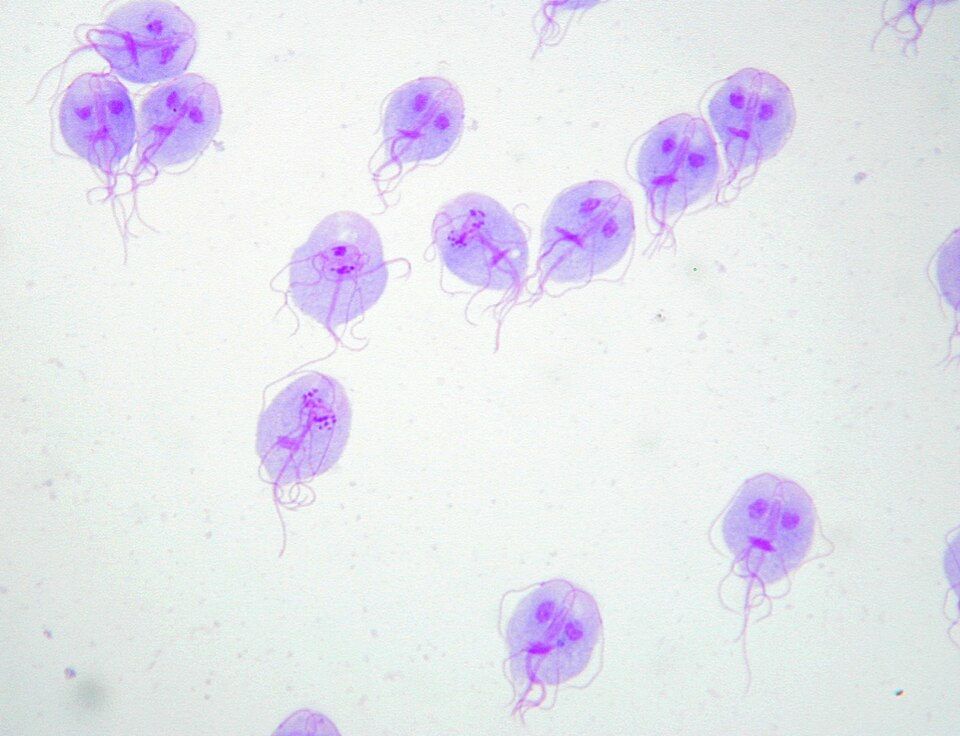
A stained faecal smear from a hiker with two weeks of greasy, foul-smelling diarrhoea shows the structure illustrated. Which organism does this pear-shaped binucleate flagellate represent?
A Gram-stained sputum smear shows numerous neutrophils containing Gram-positive lancet-shaped diplococci, with very few squamous cells. Which conclusion is supported by this finding?
Knowing where each method stops being reliable
Every microbiology method has boundaries, and the technologist has to know them before a result leaves the bench. Culture recovers only organisms that grow on the media and atmosphere chosen, so fastidious, anaerobic, intracellular and antibiotic-suppressed organisms can be missed. Rapid antigen tests trade sensitivity for speed. Nucleic acid tests detect sequence rather than viability, so they can stay positive after successful treatment. Susceptibility methods have organism and drug combinations that are not interpretable at all. Recognising these limits changes what happens next: adding a method, adding a comment, or telling the requester that a negative does not exclude the diagnosis.
A rapid antigen test for group A Streptococcus is negative on a throat swab from a child with fever, sore throat and tender cervical nodes. What is the appropriate next step?
A patient treated for Clostridioides difficile infection has a repeat stool nucleic acid test four weeks later that is still positive, although the stool is now formed and symptoms have gone. Why is this result an unreliable marker of ongoing infection?
Working a specimen through to a named organism
Identification is a sequence of decisions rather than one test. From the primary plates the technologist picks colonies that fit the clinical picture, checks purity, and applies Gram reaction, catalase, oxidase and other rapid tests to place the isolate in a group. That group sets the next step: a biochemical panel, a chromogenic medium, mass spectrometry or a molecular target. Results are read together, and a profile that conflicts with the Gram reaction or the colony appearance is re-checked rather than reported. Depth of work-up is matched to the specimen, since a sterile site is pursued much further than a superficial swab.
A urine culture yields a pure growth of a lactose-non-fermenting, oxidase-negative Gram-negative rod. Which result set points to Proteus mirabilis?
Broth from a signalling blood culture is stained and reveals clustered, purple-staining cocci; growth from the subculture is catalase positive. Which test best separates the likely pathogen from common skin commensals?
Making a slide that can actually be read
Preparation decides whether microscopy is worth doing at all. Smears are made from the purulent or otherwise representative portion of the specimen, spread thinly enough that cells lie in a single layer, air dried and fixed before staining. Thick smears trap stain and cannot be read; smears taken from the wrong part of a sputum or a swab answer the wrong question. Wet preparations for fungal elements are cleared with potassium hydroxide to dissolve keratin, while preparations for motility and protozoa are examined promptly before the organisms stop moving. Slides are labelled as they are made, never afterwards.
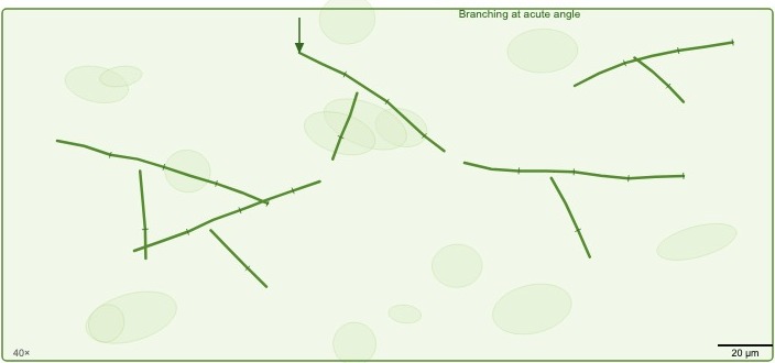
Skin scrapings from a scaling plantar lesion are mounted as shown before microscopy. What is the purpose of adding potassium hydroxide to the preparation?
An expectorated sputum is received for Gram staining and culture. Which part of the specimen should be sampled to give the most informative slide?
Handling specimens on their way to a molecular assay
Molecular testing begins at reception. The technologist checks that the specimen sits in a transport medium the assay is validated for, that stability limits have not been exceeded, and that the volume supports both the request and a repeat. Aliquots are taken in a designated clean area with dedicated equipment, using aerosol-resistant tips and one tube at a time to keep patients separate. Each aliquot is labelled at the point of transfer and frozen at the temperature the assay requires if testing is delayed. Repeated freezing and thawing degrades nucleic acid and is recorded whenever it cannot be avoided.
Several nasopharyngeal swabs in viral transport medium must be aliquoted before a molecular run. Which practice most reduces the risk of cross-contamination between patients?
Setting up amplification reactions without contaminating them
Amplification set-up is as much about containment as it is about chemistry. Work moves in one direction through separated rooms: mixes are made first, template is added next, and amplified product is read last, with dedicated coats, pipettes and filtered tips, and nothing travels backwards. The technologist assembles the master mix in the clean area, adds template last, and includes a no-template control, a positive control and an internal control in every run. Reaction volumes, primer and probe concentrations and cycling parameters follow the validated protocol, because even small changes alter sensitivity. Amplified product is the dominant contamination risk and stays in its own room.
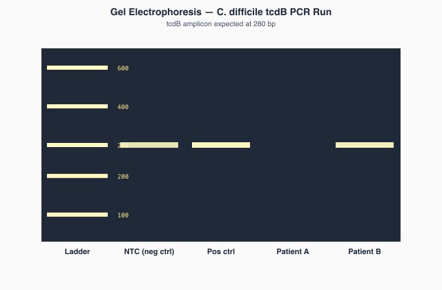
The gel shown was run at the end of a shift. The no-template control lane carries a band at the same size as the positive control band. What conclusion should the technologist draw about this run?
A laboratory validates a new real-time PCR for herpes simplex virus in cerebrospinal fluid. Which control best demonstrates that inhibitors in one individual specimen have not caused a false negative?
Choosing the media a specimen actually needs
Media selection is a clinical decision made at the bench. The technologist matches plates and broths to the specimen type, the normal flora of that site, and the organisms named or implied on the request. Non-selective media such as blood agar recover most pathogens; selective and differential media suppress background flora and separate colonies by their reactions; enrichment broths recover small numbers from sterile-site and screening specimens. Requests aimed at uncommon pathogens, including Vibrio, Yersinia, Legionella, Bordetella and Nocardia, need media that are not on the routine set-up, which is why the clinical details are read before plating.
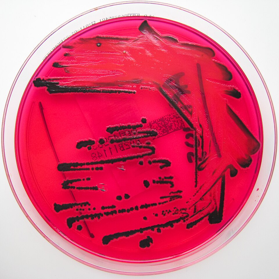
A stool specimen from a child with fever and bloody diarrhoea is plated onto the medium shown, which has produced colonies with black centres. Which property of the medium produces that appearance?
A stool culture is requested for a traveller returning from coastal Asia with profuse watery diarrhoea, and the requisition asks specifically about Vibrio. Which medium should be added to the routine enteric set-up?
Incubating cultures under the conditions the organism needs
Once plated, growth depends on atmosphere, temperature and time. Routine aerobic plates are held at 35-37 °C, chocolate agar for fastidious organisms needs added carbon dioxide, anaerobic plates need an oxygen-free environment established quickly and then left undisturbed, and Campylobacter needs a microaerophilic atmosphere at 42 °C. Some organisms simply need longer: anaerobes, Actinomyces, Brucella, fungi and mycobacteria are held for days to weeks. The technologist also protects the culture from itself, since swarming, overgrowth and drying all destroy an otherwise good plate. Incubators are monitored for temperature and, where it applies, for gas concentration.
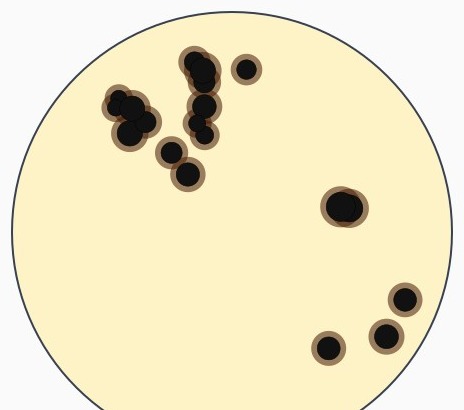
An abdominal abscess aspirate is plated on the selective medium shown and incubated anaerobically for 48 hours, producing the black colonies illustrated. Which group of organisms does this appearance identify presumptively?
A stool culture set up for Campylobacter shows no growth on the selective plate after 48 hours, while the routine plates from the same specimen grew normal enteric flora. Which incubation condition should be checked first?
Presumptive identification from the plate and a spot test
Long before a full identification is available, colony appearance plus one rapid test can narrow an isolate usefully. The technologist reads colony size, pigment, texture, odour, haemolysis and any effect on the surrounding medium, then chooses a spot test that separates the likely candidates: catalase, oxidase, coagulase, indole, PYR, bile solubility, germ tube or a rapid latex kit. These results are presumptive and are communicated as such, but they are often enough to guide immediate therapy in a serious infection. Controls are run beside each spot test, because false reactions from old growth or the wrong medium are common.
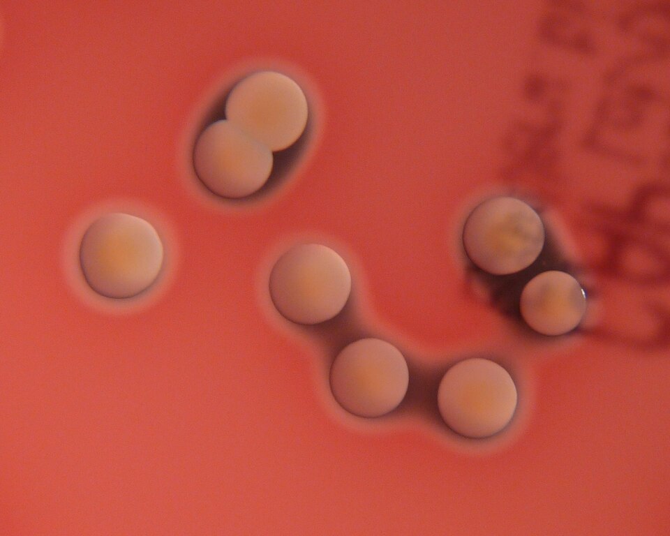
A surgical wound swab produces the golden colonies with surrounding clear zones shown after 24 hours of incubation. Which spot test result would support a presumptive report of Staphylococcus aureus?
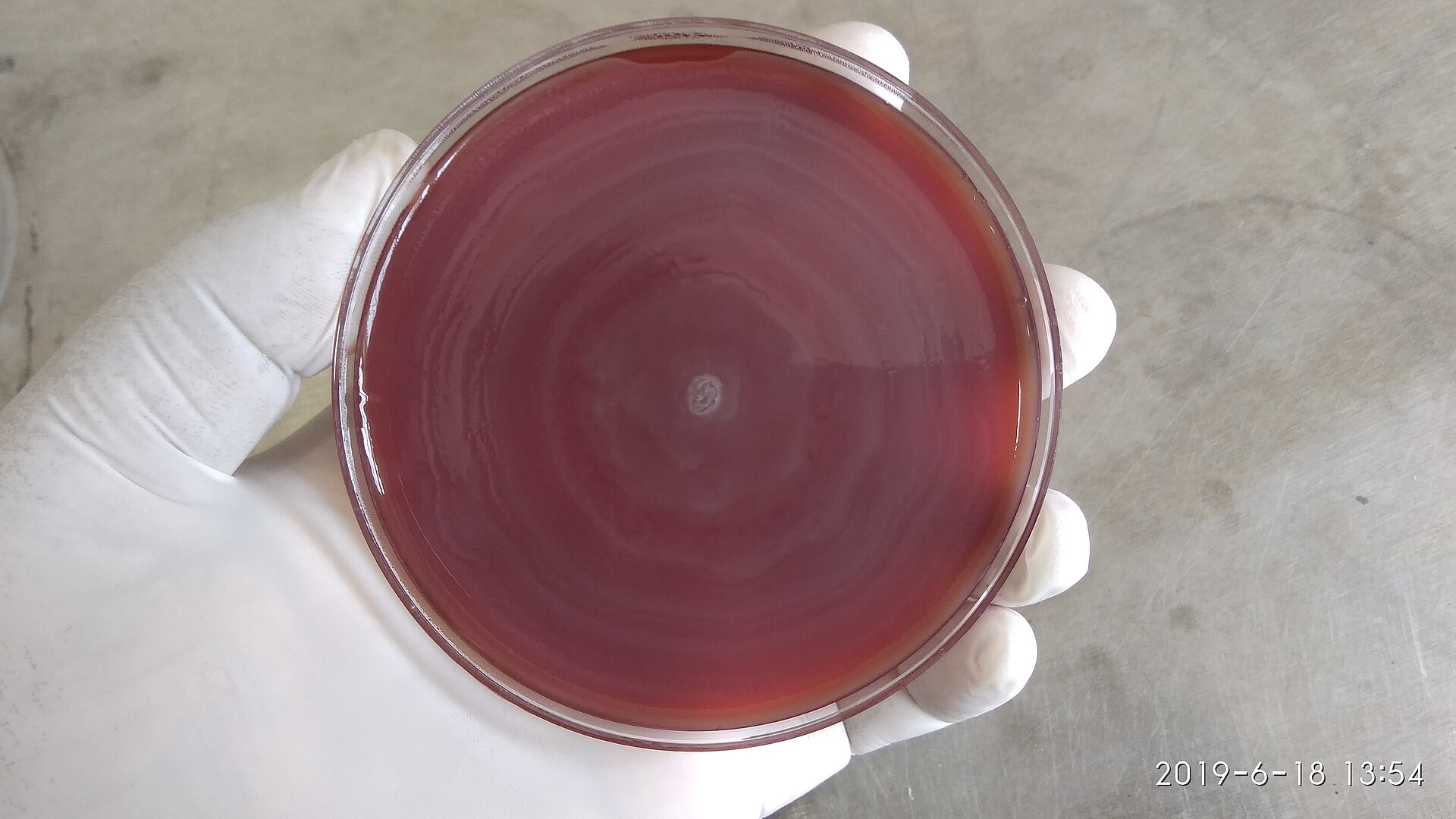
A catheter urine culture produces the spreading growth across the blood agar plate shown, and the plate has a strong ammoniacal smell. Which rapid test result would best support the presumptive identification?
Deciding whether a specimen is worth testing
A specimen that does not represent the site of infection produces a misleading result, so adequacy is judged before work proceeds. The technologist checks volume against what the requested tests consume, the container and transport medium, the time since collection, the temperature during transport and, for sputum and some other samples, a screening smear that weighs squamous cells against neutrophils. Dry swabs, leaking containers, formalin-fixed material sent for culture and specimens delayed beyond their stability window are raised with the requester. Rejection is documented with a reason, and the requester is told what to send instead.
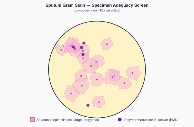
The 10× screening field shown comes from an expectorated sputum submitted for routine bacterial culture. Given the number of squamous epithelial cells present, what should the technologist do?
A single tube containing 0.3 mL of cerebrospinal fluid arrives with requests for bacterial culture, a meningitis nucleic acid panel and a cell count. What should the technologist do?
Reading the cellular and non-cellular background of a preparation
Microscopy in microbiology is not confined to organisms. The technologist assesses the inflammatory response, the host cells present and the non-cellular material: mucus, fibrin, keratin, crystals, capsular halos and stain precipitate. These observations qualify the culture result. Neutrophils with no visible organisms may point to a fastidious or partially treated infection; organisms with no inflammation may reflect colonisation; stain precipitate and drying artefact are reported as nothing at all. Capsules, spores and inclusions are shown better by a wet or negative preparation than by a Gram stain, which is why the preparation is matched to the question being asked.
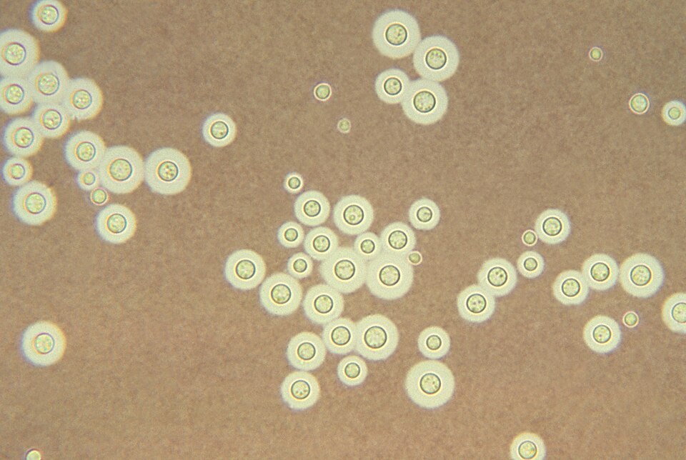
Cerebrospinal fluid sediment from a patient with advanced HIV infection is examined as shown. What does the clear zone surrounding each budding yeast cell represent?
A Gram stain of a joint aspirate shows numerous neutrophils but no organisms after the whole smear has been examined carefully. How should this finding be handled?
Grading what is seen down the microscope
Several microbiology reports are semi-quantitative, and the grade changes how the result is used. Acid-fast smears are graded from the number of bacilli seen per field or per length of smear, and that grade reflects how infectious the patient is. Gram stains are reported with scaled terms for organisms and separately for inflammatory cells. Urine microscopy counts elements per field or per volume in a counting chamber. The technologist reads at the defined magnification, examines the required number of fields, applies the published scale rather than an impression, and has a colleague confirm grades that sit near a reporting threshold.
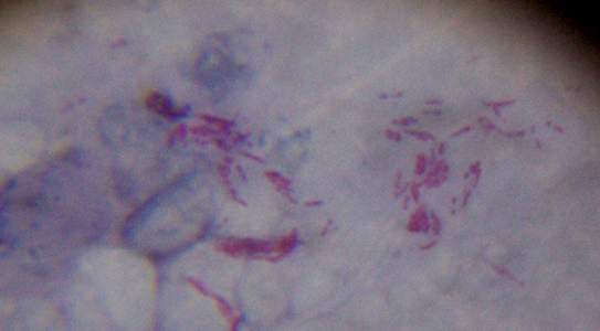
A Ziehl-Neelsen smear from a sputum specimen shows about 10 acid-fast bacilli in the field illustrated, with similar numbers in the other fields examined. How should the technologist express this finding?
Turning colonies on a plate into a reportable count
Urine and some other quantitative cultures are inoculated with a calibrated loop so that colonies can be converted into organisms per millilitre or per litre. The loop delivers a known volume and the count is divided by it, so a 1 µL loop multiplies the colony count by one thousand for a result per millilitre, or by a million for a result per litre; a 10 µL loop by one hundred and by one hundred thousand. The technologist streaks the whole loopful in the pattern the method specifies, counts each morphological type separately, and reports the count together with the identification. Counts are read against the collection method and the patient, since catheter, paediatric bag and mid-stream samples carry different thresholds, and three or more colony types usually signals contamination.
A mid-stream urine is plated with a calibrated 1 µL loop and grows about 150 colonies of a single morphology after 24 hours. By what factor is the colony count multiplied to express the result per millilitre?
A mid-stream urine from a woman with dysuria grows three different colony types, each at roughly the same count. How should this culture be reported?
Interpreting amplification data before a result is released
A molecular run is judged on its curves and its controls, not on a single call. The technologist checks that the no-template control stayed flat, that positive and internal controls amplified within their expected ranges, and that each patient curve has a genuine exponential phase rather than baseline drift or a probe artefact. Cycle threshold values are read against the validated cutoff, and values sitting close to it are repeated or reflexed to another method. Where an assay reports variants or resistance markers, only validated targets are interpreted, and an absent target is reported as not detected rather than as absence of disease.
In a qPCR run, the no-template control is flat, the positive control amplifies as expected, and one patient sample crosses the threshold at cycle 34 with a clean exponential curve. What action does this pattern support?
In a multiplex gastrointestinal panel, one specimen shows no amplification of its internal control while every batch control passes. What should the technologist do?
Getting nucleic acid out of a specimen intact
Molecular results are only as good as the extract behind them. The technologist selects an extraction chemistry that suits the matrix, whether that is swab transport medium, whole blood, stool, a respiratory sample or fixed tissue, and follows the lysis, binding, washing and elution steps without shortcuts. Inhibitors are the main enemy: haem, heparin, mucin, faecal polysaccharides and residual ethanol all suppress amplification, and each is handled by a specific wash or dilution step. Nucleases are controlled by working quickly, keeping extracts cold and using nuclease-free consumables. An internal control carried through extraction shows whether the process worked for that specimen.
A whole blood specimen submitted for a viral load assay was collected into a heparin tube. Why is this tube unsuitable for the molecular request?
Microscopic examination of urine in the microbiology laboratory
Urine microscopy supports and sometimes redirects the culture. The technologist examines a standardised volume, either uncentrifuged in a counting chamber or as a resuspended sediment, and reports leukocytes, erythrocytes, epithelial cells, bacteria, yeast and casts against the volume examined. Squamous cells and several bacterial morphologies point to contamination and predict a mixed culture. Leukocytes with no growth raise sterile pyuria, which prompts thought about partially treated infection, fastidious organisms, mycobacteria or a non-infectious cause. Microscopy is read alongside the colony count, and any disagreement between the two is investigated rather than reported unresolved.
A mid-stream urine shows 50 leukocytes per high-power field on microscopy, but the culture is sterile after 24 hours of incubation. Which follow-up best addresses this pattern?
A urine sediment shows numerous squamous epithelial cells together with several different bacterial morphologies. What does this microscopy predict about the culture?
Testing an isolate against the antimicrobials that matter
Susceptibility testing is valid only when the method is followed exactly. The technologist prepares the inoculum to a defined turbidity from fresh, pure growth, plates it within the specified time, applies disks or a gradient strip firmly, and incubates at the stated agar depth, temperature and atmosphere. Zones are measured at the point of complete inhibition and read against current interpretive criteria for that organism and drug combination. The panel is chosen for the organism, the specimen site and the local formulary, and every set of results is screened for patterns that make no biological sense before it is released.
A disk diffusion plate was set up from a suspension prepared by eye, without comparison against a turbidity standard, and the suspension was clearly too heavy. What effect does an over-dense inoculum have on the zones?
A Staphylococcus aureus isolate from a blood culture tests susceptible to oxacillin but resistant to cefoxitin. What should the technologist do before releasing the report?
Keep reading with a free account
The rest of this page — 19 more sections and 35 practice questions — is free to read with an MLTPrep account.
No card required.
Back to all clinical microbiology areas in the Clinical Microbiology study guide.
MLTPrep is an independent study resource. Not affiliated with or endorsed by CAMLPR. Always confirm current requirements with CAMLPR directly.